Foot Injuries in the ED
/
BACKGROUND
Foot injuries are common and can have significant implications for a patient’s well-being, functional capacity, and finances. Foot injuries can negatively impact quality of life and ability to work both in the immediate post-injury period (e.g., due to pain, weight bearing status, discomfort) and potentially later due to any injury sequelae (e.g., nonunion, post-traumatic arthritis or deformity). At a larger level, foot and ankle surgeries are responsible for more than 11 billion dollars spent annually just within the Medicare population [1].
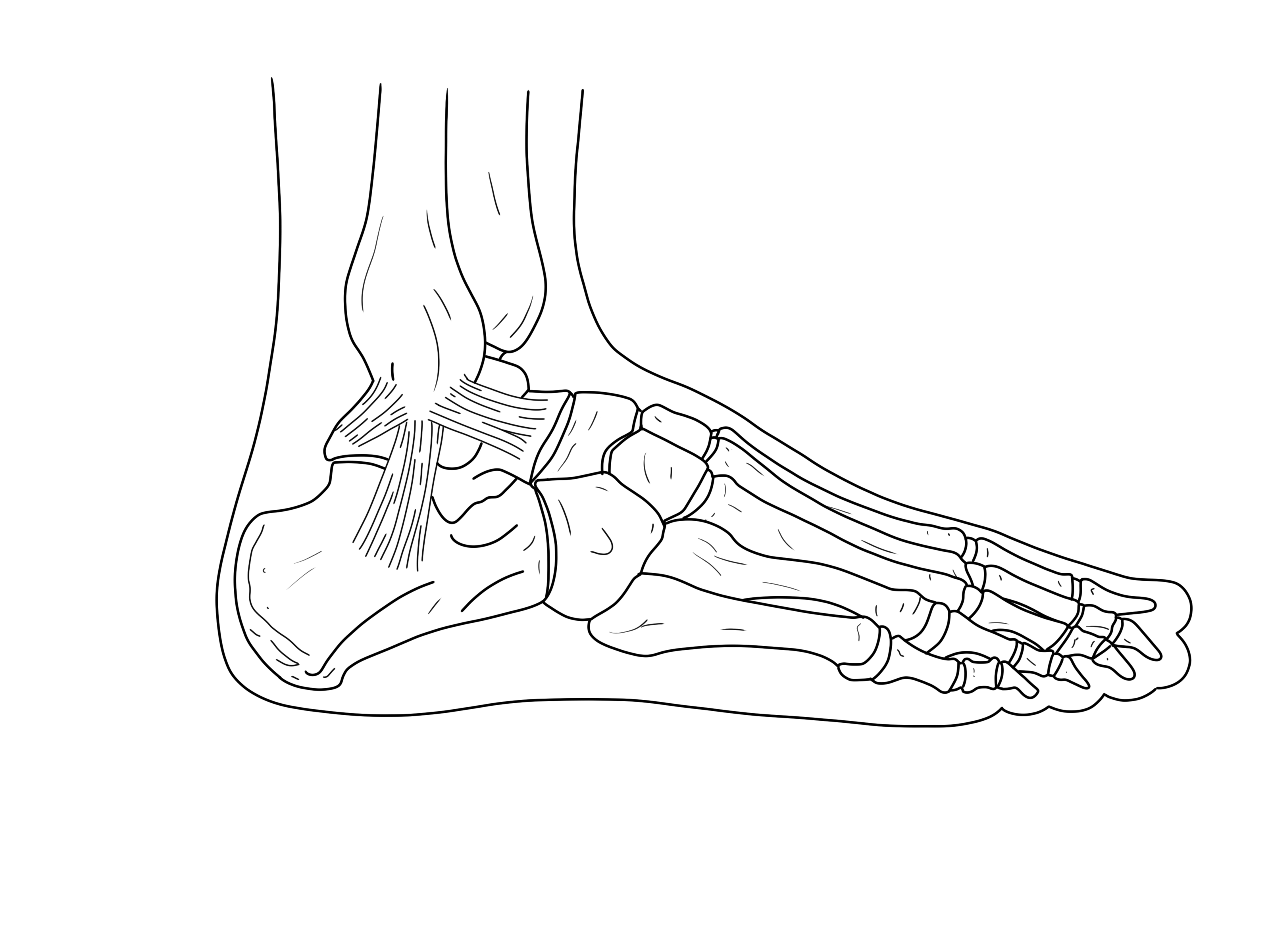
As an EM physician, it is important to have an understanding of the spectrum of foot injuries and how these are appropriately evaluated. Certain injuries carry risks of further injury, injury-related complications, and poor outcomes which are exacerbated if they are inappropriately managed in the ED. This post will cover some of the most common and important injuries, but is not comprehensive. Injuries discussed are shown in Image 1.
Foot and ankle injuries commonly co-occur. For discussion of ankle injuries, see our previous post.

Figure 1 - Common sites of foot injuries - adapted from https://upload.wikimedia.org/wikipedia/commons/3/37/Ankle-lateral.png
APPROACH TO FOOT INJURIES
Presentation and Mechanism
Foot injuries most often occur from a traumatic force exerted on a foot in a non-neutral position, but can also occur from a variety of mechanisms including polytrauma, MVCs, crush injuries, GSWs, and long-term stress or overuse. Certain injuries are classically associated with certain mechanisms or degrees of traumatic impact (e.g., fractures of the talar body, talar neck, and calcaneus require high-impact trauma). Stress fractures are caused by repetitive microtrauma and stress on the bone, and therefore present with more subacute or chronic symptoms. Unstable injuries may be obvious at time of presentation, with the patient not being able to bear any weight. While knowing the mechanism can provide clues to potential injuries or increase your index of suspicion for certain injuries, mechanisms of injuries can be variable. Respect, but do not rely upon, the mechanism.
Exam
The exam (along with the history) is critical for proper diagnostic evaluation. Use your exam to guide your initial management as well as your imaging decisions and differential diagnosis.
Any significant bleeding should first be controlled in the setting of an open injury
Neurovascular assessment (perfusion, pulses, sensory function, motor function) should be prioritized, regardless of acuity. Abnormal neurovascular findings suggest injury to nearby structures (nerves and blood vessels) from dislocations, bony trauma, or increased compartment pressures.
Gross deformities often suggest fracture or dislocation.
Skin tenting or other overlying skin changes are often indicative of impending open fracture or risk of skin necrosis. Tongue-type calcaneal fractures are at particular high risk of skin necrosis [2]. LisFranc injuries have a characteristic pattern of plantar ecchymosis [3].
Palpate all bones and joints. In general, consider imaging of joints adjacent to an area of bony tenderness and imaging of bones adjacent to areas of joint tenderness.
Range all joints. If active ROM is impaired, perform passive ROM. Impaired ROM can be indicative of dislocation, ligamentous injury, or intra-articular pathology.
Certain provocative tests or inability to bear weight can be clues to instability. If instability is suspected or apparent, avoid having the patient bear weight.
In the setting of a more diffuse traumatic injury, always examine all other bones and joints even if the patient is only complaining of pain at the foot (their foot injury may be a distracting injury).
Consider examination and diagnostic evaluation for any injuries that are likely to co-occur (e.g., calcaneal fractures associated with contralateral calcaneal fracture, pilon, or vertebral compression fracture [2]).
Appropriate Imaging
The vast majority of foot injuries require at least basic foot radiographs for proper evaluation, and often ankle films as well. Obtain at least 3 views when possible. While the Ottawa Foot Rules [4] are often referenced and tested on exams, these criteria can miss significant injuries. Special views can help identify certain fractures on x-rays (e.g., axial view for calcaneal fractures [2], Canale view for talar fractures [5]). Stress films are useful in assessing LisFranc injuries. Some fractures are difficult to see on XRs, even with special views, so consider CT if your index of suspicion is high. Certain injuries require CTs for further characterization or surgical planning. MRI can be used in place of CT.
Management
Immediate management
Control significant bleeding (e.g., direct pressure, tourniquet)
Address any impairments in neurovascular status (e.g., role of consulting services, emergent closed reduction, need for vascular imaging).
In the ED
Pain control, including consideration for nerve blocks in certain injuries
Wound management (irrigation and debridement, possible closure, consideration of Abx, tetanus immunization)
Closed reduction and/or splinting (make sure to obtain repeat XRs after)
Determine if injury is operative vs. non-operative
Determine if Orthopedics (or other specialist) consultation is needed
Calcaneal or talar fracture
Unstable or surgical injury (or if this is suspected)
Uncertainty in diagnosis
Uncertainty in management (e.g., weight-bearing status, operative vs. non-operative)
If necessary/helpful to ensure adequate follow-up
At time of discharge
Counsel on expected course of injury, supportive care (e.g., RICE), and symptomatic management (e.g., NSAIDs, Tylenol, opioid pain medication)
Counsel on ER return precautions, complications that are more likely in certain injuries (e.g., DVT, compartment syndrome), and consider prophylactic measures as appropriate
Brace, other cast, walking boot, hard-sole shoe, etc.
Crutches and crutch training
Give clear instructions on weight-bearing status and use of any braces or walking boots given
Ensure there is a clear follow-up plan and provide any necessary referrals
Who they should see (e.g., primary care physician, Orthopedics, other specialist)
Whether they need to be seen definitely or only if needed, and when
Confirm correct patient contact information in EMR
Open Fractures and Injuries
Wounds that are not open fractures should receive irrigation and debridement, closure (if indicated), tetanus vaccination, consideration of antibiotics, and wound care instructions. Orthopedics should be consulted for any wounds near joints with concern of extension into the joint (i.e., traumatic arthrotomy), with a lower threshold to consult if the joint is non-native.
Open fractures and dislocations are generally managed with bleeding control, close assessment of neurovascular status, antibiotics with routine gram-positive coverage (Ancef; clindamycin if penicillin allergy), tetanus vaccination, and early irrigation and debridement in the ER with low pressure normal saline [6]. Open fractures can be categorized according to the Gustilo-Anderson classification system [7], though this has been critiqued for poor interrater reliability. Smaller wounds may not require antibiotics, but if in doubt, one dose of Ancef in the ED is unlikely to cause significant harm. Larger wounds (Gustilo-Anderson Type III) should also receive gram-negative microbial coverage. Wounds with potential soil or fecal contamination should receive high-dose penicillin [6]. The management of open fractures is further discussed in these TamingTheSRU posts:

Table 1 A - Management considerations for lisfranc injuries and calcaneal fracture

Table 1 B - Management considerations for 5th metatarsal fractures

Table 1C: Management considerations for talar fractures
References
Belatti DA, Phisitkul P. Economic burden of foot and ankle surgery in the US Medicare population. Foot & Ankle Int. 2014,35(4):334-340. doi: 10.1177/1071100713519777
Forsthoefel C. Calcaneus fractures. [Updated 2022 Nov 23]. In: OrthoBullets [Internet]. Available from: https://www.orthobullets.com/trauma/1051/calcaneus-fractures
Alrasheedi A, Koyfman A, Alerhand S. Foot injuries in the emergency department. [2015 Apr 28]. In: EM Docs [Internet]. Available from: http://www.emdocs.net/foot-injuries-in-the-emergency-department/
Pires R, Pereira A, Abreu-E-Silva G, Labronici P, Figueiredo L, Godoy-Santos A, Kfuri M. Ottawa ankle rules and subjective surgeon perception to evaluate radiograph necessity following foot and ankle sprain. Ann Med Health Sci Res. 2014 May;4(3):432-5. doi: 10.4103/2141-9248.133473. PMID: 24971221; PMCID: PMC4071746.
Weatherford B. Talar neck injuries. [Updated 2022 May 14]. In: OrthoBullets [Internet]. Available from: https://www.orthobullets.com/trauma/1048/talar-neck-fractures
Fares A, Szatkowski J. Tibial plafond fractures. [Updated 2023 Apr 22]. In: OrthoBullets [Internet]. Available from: https://www.orthobullets.com/trauma/1046/tibial-plafond-fractures
Pires R, Pereira A, Abreu-E-Silva G, Labronici P, Figueiredo L, Godoy-Santos A, Kfuri M. Ottawa ankle rules and subjective surgeon perception to evaluate radiograph necessity following foot and ankle sprain. Ann Med Health Sci Res. 2014 May;4(3):432-5. doi: 10.4103/2141-9248.133473. PMID: 24971221; PMCID: PMC4071746.
Radiopaedia. Calcaneal fracture. [Updated 2023 Mar 30]. Available from: https://radiopaedia.org/articles/calcaneal-fracture?lang=us
Aiyer A, Moore DW. Talus fracture (other than neck). [Updated 2023, Apr 14]. In: OrthoBullets. [Internet]. Available from: https://www.orthobullets.com/trauma/1049/talus-fracture-other-than-neck
Wong PK, Hanna TN, Shuaib W, Sanders SM, Khosa F. What's in a name? Lower extremity fracture eponyms (Part 2). Int J Emerg Med. 2015 Dec;8(1):76. doi: 10.1186/s12245-015-0076-1. Epub 2015 Jul 25. PMID: 26223985; PMCID: PMC4512960.
Steffes MJ, Weatherford M. 5th metatarsal base fractures. [Updated 2023 Feb 3]. In: OrthoBullets [Internet]. Available from: https://www.orthobullets.com/foot-and-ankle/7031/5th-metatarsal-base-fracture
Authorship
Written by: Isabel Lott, MD, PGY-1, University of Cincinnati Department of Emergency Medicine
Peer Review by Bret Betz, MD, Associate Professor, University of Cincinnati Department of Emergency Medicine
Editing and Posting by Jeffery Hill, MD MEd, Associate Professor, University of Cincinnati Department of Emergency Medicine
CITE AS
Lott, I., Betz, B., Hill, J. (May 23, 2023) Foot Injuries in the ED. TamingtheSRU. https://www.tamingthesru.com/blog/diagnostics/foot-injuries-in-the-ed


