Ultrasound of the Month: Peritonsillar Abscess
/THE CASE
A female in her 20’s presented to the emergency department with concern for oral swelling. She had previously been evaluated at an outside facility for sore throat and was subsequently diagnosed with acute pharyngitis and left tonsillar hypertrophy. She was placed on a course of antibiotics and sent home, however, she continued to have worsening throat pain and edema of her left tonsil with associated difficulty swallowing. She endorsed low grade fevers and chills as well as odynophagia, dysphagia, and some shortness of breath.
On examination, the patient was noted to have asymmetric swelling of the left tonsillar region with no uvular deviation. There was no trismus. The remainder of her exam was unremarkable. Lab work showed significant leukocytosis of 24.5. A bedside ultrasound was performed, and the adjacent images were obtained:
clip 1 demonstrates the presence of an anechoic/hypoechoic, heterogeneous collection by the left tonsil
clip 2 similarly to clip 1, the same structure can be seen in a different plane
clip 3 demonstrates a submandibular view of the abscess
clip 4 demonstrates the use of color doppler to identify the carotid artery, which can be seen in the far field in this clip
PERITONSILLAR ABSCESS PATHOLOGY
A peritonsillar abscess is a walled off collection of pus that develops in the peritonsillar space, a space that exists between the tonsillar capsule and the superior constrictor muscle. As the pus accumulates and the abscess grows, patients may experience uvular deviation or in severe cases, airway concern. Most peritonsillar abscesses are polymicrobial and often contain common oropharyngeal microflora. They represent the most common type of deep neck infection and are often characterized by a severe sore throat, odynophagia with drooling, trismus, and muffled voice (1). Although rarely fatal, peritonsillar abscess can carry significant morbidity if not identified and treated in a timely manner. Severe complications of peritonsillar abscesses include infection spreading to additional deep spaces of the neck as well as mediastinitis (2).
IMAGING WITH ULTRASOUND
Peritonsillar abscess is one of the most common deep space infections of the head and neck contributing significantly to health care costs in the United States. It is estimated that peritonsillar abscess costs approximately 150 million in health care expenditure yearly (3). One factor contributing to this increased cost is the increased usage of computed tomography (CT) in the emergency department to confirm the diagnosis prior to incision and drainage. The use of point of care ultrasound for diagnosis and guidance of treatment may be an adequate alternative to obtaining CT imaging. Studies have shown that intraoral ultrasound can accurately diagnose peritonsillar abscesses and reduce the risk of blind needle aspirations (4). An additional study found that intraoral ultrasonography has a sensitivity of 100% for ruling out peritonsillar abscess making it a strong initial imaging modality and rendering CT imaging unnecessary in the majority of patients (5).
INTRAORAL APPROACH

Image 1 demonstrates a measurement of 1.6cm from the mucosal surface to the center of the abscess. This can be helpful when performing drainage to avoid deep insertion of the needle
The intraoral or transoral approach is the most common approach. A prospective randomized study found that clinician diagnostic accuracy at diagnosing PTA when using intraoral ultrasound was 100% versus landmark needle aspiration which was 64% (6). Patient preparation is one of the most important aspects of successful intraoral ultrasound. The patient should be sitting upright and the oropharynx should be prepared by using a topical anesthetic that is sprayed liberally. Intraoral ultrasonography is performed using an intracavitary transducer covered with a sterile sheath. The transducer will come in contact with tonsillar tissue as peritonsillar abscesses usually form within a potential space. It can be helpful to have the patient hold the probe itself and place it in the area of maximum discomfort. Once the area of interest is identified it should be scanned in two orthogonal planes. The use of intraoral ultrasonography is not only helpful for the confirmation of an abscess, but can also inform the provider on the depth and location of the carotid artery in relation to the fluid collection, as seen in image 1.
SUBMANDIBULAR APPROACH

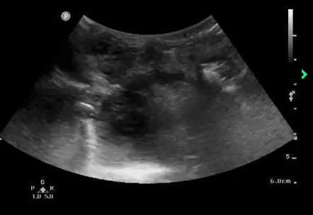
Image 2 Demonstrates the ideal placement of the probe to identify the tonsil and abnormalities surrounding it
Patients with a PTA can often experience significant trismus. This is usually due to the associated inflammation of the adjacent pterygoid muscles (7). The presence of trismus can lead to challenges when performing intraoral ultrasonography. Evidence regarding use of steroids is controversial but some studies have shown steroids lead to improvement in trismus. Methylprednisolone 125mg intravenously (IV) or dexamethasone 10mg orally (PO) or intramuscularly (IM) is typically recommended for treatment (8). However, the peak effect of steroids is usually not immediate. If the patient is still unable to tolerate intraoral ultrasonography despite medication and coaching, the submandibular approach can be considered. This can be performed with the use of a high frequency linear probe as seen in image 2. The patient can be supine or upright and the probe should be placed in the submandibular space, parallel to the mandible . Few studies have explored the specificity and sensitivity of this approach, but case reports have been published and describe its success in assisting with diagnosis (9). The image below demonstrates the presence of an anechoic region adjacent to the right tonsil when compared to the left, indicative of a right sided PTA–which was successfully drained after diagnosis with this approach (9).
MANAGEMENT
Definitive treatment for peritonsillar abscess includes incision and drainage. Once a fluid collection in the peritonsillar space is confirmed, the provider can perform drainage via needle aspiration or a small incision can be made into the area of interest. Once drainage has been performed, the patient should be placed on antibiotics. Refer to Mastering Minor: Peritonsillar Abscess for tips and tricks regarding management!
CASE RESOLUTION
The patient underwent incision and drainage of the confirmed left peritonsillar abscess in the emergency department. During this procedure, a moderate amount of purulent fluid was drained. The patient was given a dose of dexamethasone and one dose of intravenous antibiotics and ultimately discharged home with oral antibiotics and outpatient follow up with otolaryngology.
AUTHORED BY Jazmyn shaw, MD
Dr. Shaw is a PGY-3 resident in Emergency Medicine at the University of Cincinnati and a Resident Editor of Ultrasound of the Month.
PEER REVIEW BY Patrick Minges, MD
Dr. Minges is an Ultrasound Fellowship trained Assistant Professor in Emergency Medicine at the University of Cincinnati.
EDITING AND LAYOUT BY MARTINA DIAZ MCDERMOTT, MD
Dr. Diaz is a PGY-4 resident in Emergency Medicine at the University of Cincinnati and the current Resident Editor of Ultrasound of the Month.
REFERENCES
El-Saied S, Puterman M, Kaplan DM, Cohen-Lahav M, Joshua BZ. Involvement of minor salivary glands in the pathogenesis of peritonsillar abscess. Otolaryngol--Head Neck Surg Off J Am Acad Otolaryngol-Head Neck Surg. 2012;147(3):472-474. doi:10.1177/0194599812445552
Matsuda A, Tanaka H, Kanaya T, Kamata K, Hasegawa M. Peritonsillar abscess: a study of 724 cases in Japan. Ear Nose Throat J. 2002;81(6):384-389.
Johnson RF, Stewart MG. The contemporary approach to diagnosis and management of peritonsillar abscess. Curr Opin Otolaryngol Head Neck Surg. 2005;13(3):157-160. doi:10.1097/01.moo.0000162259.42115.38
Buckley AR, Moss EH, Blokmanis A. Diagnosis of peritonsillar abscess: value of intraoral sonography. AJR Am J Roentgenol. 1994;162(4):961-964. doi:10.2214/ajr.162.4.8141026
Nogan S, Jandali D, Cipolla M, DeSilva B. The use of ultrasound imaging in evaluation of peritonsillar infections. The Laryngoscope. 2015;125(11):2604-2607. doi:10.1002/lary.25313
Costantino TG, Satz WA, Dehnkamp W, Goett H. Randomized trial comparing intraoral ultrasound to landmark-based needle aspiration in patients with suspected peritonsillar abscess. Acad Emerg Med Off J Soc Acad Emerg Med. 2012;19(6):626-631. doi:10.1111/j.1553-2712.2012.01380
Paul E, Toldt C, Della Rosa A. An Atlas of the Human Anatomy for Students and Physicians: Volume 1. 1919
Battaglia A, Burchette R, Hussman J, Silver MA, Martin P, Bernstein P. Comparison of Medical Therapy Alone to Medical Therapy with Surgical Treatment of Peritonsillar Abscess. Otolaryngol Head Neck Surg. 2018;158(2):280-286. doi:10.1177/0194599817739277
Rehrer M, Mantuani D, Nagdev A. Identification of peritonsillar abscess by transcutaneous cervical ultrasound. The American Journal of Emergency Medicine. 2013; 31(1):267.e1-3. doi: 10.1016/j.ajem.2012.04.021.


