Ultrasound of the Month: Flexor Tenosynovitis
/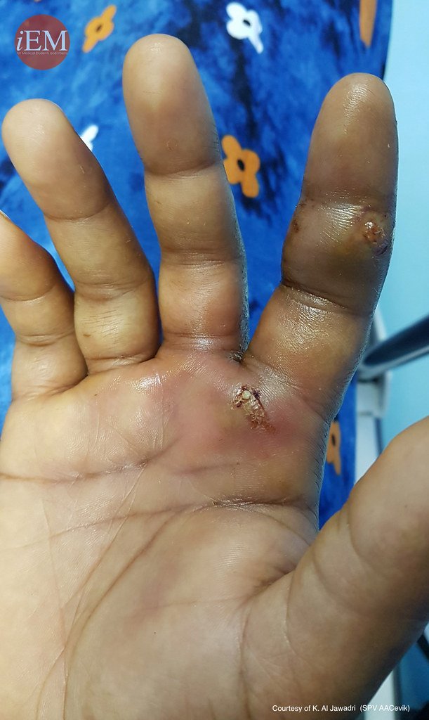
Figure 1. Sample image of findings seen in flexor tenosynovitis (8)
The Case
A 44-year-old man presents to the emergency department for pain localized to the fourth digit of his left hand. The patient has a past medical history significant for Intravenous Drug Use (IVDU) with multiple prior presentations for soft tissue infections including Methicillin-resistant Staphylococcus aureus (MRSA). Additional medical history includes hepatitis C, hepatitis B, and schizoaffective disorder. He states that has had finger pain for two days and last injected heroin yesterday though not in the vicinity of the pain described today. There was no preceding trauma. He has taken no medications to treat his condition and has sought no prior treatment. With ongoing discomfort today, he called paramedics who found him hemodynamically stable, afebrile, and transported him for evaluation.
Upon evaluation he is alert and conversant, in little distress. Focused examination of the left hand reveals a warm, erythematous, and swollen fourth digit. The patient maintains the digit in a flexed position and experiences exquisite pain when attempts are made to passively extend or palpate discrete regions of the finger. He refuses to attempt active extension. The digit is neurovascularly intact. There are no other acute abnormalities identified on the remainder of the hand examination.
Figure 2. Longitudinal view of POCUS
The patient’s presentation raises concern for flexor tenosynovitis. Other differential items may include cellulitis, abscess, small joint septic arthritis, gout, and undisclosed trauma. Consultation to the orthopedic hand service is initiated and consideration is given to imaging modalities which may support the primary diagnosis including ultrasound. Point-of-care Ultrasound (POCUS) of the digit is obtained as shown in Figures 2-4. Additional workup includes labs and plain film x-ray, which reveals diffuse swelling of the fourth digit without other abnormalities. Labs are remarkable for CRP to 61 mg/dL and ESR to 33 mm/hr.

Figure 3. Longitudinal view of POCUS

Figure 4. Transverse view of POCUS
Flexor Tenosynovitis
Tenosynovitis describes an inflammatory condition involving the synovial space within a tendon sheath. This condition can result in permanent damage and dysfunction of the involved tendon and associated structures. Noninfectious causes of tenosynovitis include autoimmune disease, overuse of the digit, and idiopathic causes. Infectious causes of tenosynovitis can be particularly debilitating, developing rapidly over days. The most common isolated organism in infectious cases is staph aureus including MRSA and the mechanism of inoculation includes both direct injury and hematogenous spread (1). In the hand, this condition is estimated to represent 2.5-9.4% of all infections (2-3). The Kanavel signs provide the classic clinical picture for infectious flexor tenosynovitis.(5) These include the following four findings:
Fusiform swelling of the affected finger
Tenderness along the course of the tendon sheath
Digit held in semi-flexed posture
Pain on passive extension of the affected digit
The test characteristics for the Kanavel signs favor sensitivity over specificity and are cited at 91-97% and 51-69% respectively (6). Imaging modalities including plain film x-ray and CT scan add little to the evaluation. Ultrasound, can be quite useful in aiding providers with making this diagnosis. Its test characteristics are at least comparable to the Kanavel signs and are cited in one study as a sensitivity of 94% and specificity of 65% (7).
Tips for Musculoskeletal ultrasound of the hands / feet

Figure 5. The hand is submerged in a water bath and the probe itself can rest on the surface of the bath, without making contact with the hand
Obtaining high quality musculoskeletal ultrasound studies of the hands and feet can be challenging, particularly if the patient has a painful condition such as a soft tissue infection. As there is little tissue covering the region of interest, it is advisable to use a high frequency linear probe to optimize visualization of structures at 1-2 cm of depth. As with any ultrasound examination, images should be evaluated in both longitudinal and transverse planes. The patient will benefit from enteral or parenteral analgesia or regional anesthesia to facilitate their care. Immersing the extremity in a water bath can be extremely helpful and may allow ultrasound examination without direct contact of the ultrasound probe on the tissues as in Figure 5. The probe itself can rest at the surface of the water while the digit is submerged. In the case described here, a generous collection of ultrasound gel allowed examination of the finger with little discomfort to the patient. As shown in Figures 2-4 abnormalities concerning for tenosynovitis here included a hypoechoic fluid collection tracking along the flexor tendon just shallow to the bony cortex in the far field of the images as well as a cobblestone appearance of the subcutaneous tissues in the near field.
Management
Historically, infectious tenosynovitis of the flexor tendons of the fingers have been managed with open operative debridement. However, less invasive techniques have been developed involving catheter directed irrigation and the addition of parenteral antibiotics have significantly reduced the overall morbidity of this disease (4).
Case Conclusion
The ultrasound images are reviewed with the consulting orthopedic hand service and strongly support a diagnosis of flexor tenosynovitis. Given this, the patient is placed on IV antibiotics and admitted for operative intervention. During operative exploration, purulent fluid was obtained upon puncture of the flexor tendon sheath and sent for culture. The culture returned positive for MRSA. The patient completed a six-week course of IV vancomycin and had a complete recovery.
AUTHORED BY Carl Goff, MD
Dr. Goff is a PGY-4 in Emergency Medicine at the University of Cincinnati
PEER REVIEW BY JESSICA BAEZ, MD
Dr. Baez (@blonde_doctr) is an Assistant Professor of Emergency Medicine at the University of Cincinnati and Assistant Residency Director.
EDITING AND LAYOUT BY ARTHUR BROADSTOCK, MD AND MARTINA DIAZ, MD
Dr. Broadstock (@BroadstockMD) is a PGY-4 in Emergency Medicine at the University of Cincinnati and former Resident Editor of Ultrasound of the Month.
Dr. Diaz (@martina_diazb) is a PGY-3 in Emergency Medicine at the University of Cincinnati and the current Resident Editor of Ultrasound of the Month.
References
Ray G, Sandean DP, Tall MA. Tenosynovitis. [Updated 2021 Jul 26]. In: StatPearls [Internet]. Treasure Island (FL): StatPearls Publishing; 2022 Jan-. Available from: https://www.ncbi.nlm.nih.gov/books/NBK544324/
Giladi AM, Malay S, Chung KC. A systematic review of the management of acute pyogenic flexor tenosynovitis. J Hand Surg Eur Vol. 2015 Sep;40(7):720-8. doi: 10.1177/1753193415570248. Epub 2015 Feb 10. PMID: 25670687; PMCID: PMC4804717.
Pang HN, Teoh LC, Yam AK, et al. Factors affecting the prognosis of pyogenic flexor tenosynovitis. J Bone Joint Surg Am. 2007;89:1742–8.
Weinzweig N, Gonzalez M. Surgical infections of the hand and upper extremity: a county hospital experience. Ann Plast Surg. 2002;49:621–7.
Kanavel A. The symptoms, signs and diagnosis of tenosynovitis and major facial space abseceses. 6 ed Lea & Febiger; 1933
Kennedy CD, Lauder AS, Pribaz JR, Kennedy SA. Differentiation Between Pyogenic Flexor Tenosynovitis and Other Finger Infections. Hand (N Y). 2017 Nov;12(6):585-590.
E. Jardin, M. Delord, S. Aubry, F. Loisel, L. Obert. Usefulness of ultrasound for the diagnosis of pyogenic flexor tenosynovitis: a prospective single-center study of 57 cases. Hand Surg Rehabil, 37 (2018), pp. 95-98
Image from: Emergency Medicine Clinical Images and Videos. CC 2.0


