Twists and Turns: Identifying Maisonneuve Fractures in the ED
/Musculoskeletal injuries are a common occurrence, representing a substantial number of Emergency Department visits on an annual basis. In fact, it’s been estimated that approximately 20% of all ED visits are musculoskeletal in origin. (1) Oftentimes, Emergency Physicians are the first provider patients encounter after an injury. This places a significant responsibility on the EM physician to diagnose and treat fractures. Specifically, EM physicians should be able to recognize fractures that will likely require operative management and facilitate close follow up, such as a Maisonneuve fracture. A Maisonneuve fracture is a specific ankle fracture pattern that involves the medial malleolus, syndesmosis and proximal fibula. It can be easily missed if a provider does not routinely evaluate the proximal fibula as part of their ankle examination, as x-rays of the ankle can often appear normal. Disruption of these structures yields an unstable ankle fracture, thus making close follow up for operative management imperative. It is key to identify this fracture when patients present to the Emergency Department with ankle injuries to ensure definitive management and prevention of complications down the line.
Case
You are working solo coverage in a community hospital during a busy evening shift in the ED. You finally have time to sit down after seeing a bolus of patients that included a complex lac repair from a dog bite and a patient who required intubation for hypoxic respiratory failure. You notice a patient check in for ankle pain. Thrilled to see a patient with a potentially straightforward presentation, you hop into the room and find a 37 y.o. M with right ankle pain and swelling. The patient states he was playing a pick up basketball game with his friends one hour prior when he went up for a rebound and “landed on his ankle funny”. He states he may have heard a pop, noticed immediate swelling on the inside of his right ankle with significant pain and had difficulty moving his ankle. He was unable to bear weight and needed assistance off the court. He was able to find some old crutches and came in for an x-ray.
The patient’s physical exam is notable for significant edema along the medial malleolus of the right ankle. Tender to palpation. Limited dorsiflexion/plantar flexion secondary to pain. Sensation intact to light touch. 2+ right DP and PT pulses. Able to wiggle toes. Tender to palpation of the right fibular neck. You give the patient ibuprofen and order an x-ray.
X-ray
Figure 1: Arrows denote widening of the medial clear space and a spiral fracture of the proximal fibula.
You read your x-ray and note the patient has a proximal fibula fracture and what appears to be widening of the ankle joint. You decide to look up information for definitive weight bearing recommendations and realize the patient has a Maisonneuve fracture. Before returning to the patient’s room, you do a little more digging into what constitutes a Maisonneuve fracture and best treatment plans.
Injury
A Maisonneuve fracture is a specific type of ankle fracture accounting for approximately 7% of all ankle fractures. (2) It is a specific type of a Weber C ankle fracture. Weber C ankle fractures are characterized by ankle injuries involving the proximal fibula above the ankle mortise. The injury pattern of a Maisonneuve fracture involves a spiral fracture of the proximal fibula with associated rupture of the tibiofibular syndesmosis and the anterior fibers of the deltoid ligament. Among 41 patients presenting to an ED with an identified Maisonneuve fracture, 74% had an associated medial malleolus fracture. 24.3% of patients had a deltoid ligament injury confirmed by MRI. (3) The injury pattern associated with this type of fracture involves forced pronation-external rotation. There are different stages categorizing this mechanism of injury, Maisonneuve fractures often are often Stage III/IV as injuries to the deltoid ligament and/or medial malleolus tend to be severe. Figure 1 highlights the mechanism of injury and range of severity.
From Dr. Sachin Kale, Dr. Pratik Tank, Dr. Rahul Ghodke, Dr. Pankaj Singh, Dr. Abhiraj Patel. Functional outcome of bimalleolar ankle fractures treated with fibular plate for lateral malleolus and C.C. screws for medial malleolus. Int J Orthop Sci 2020;6(1):1126-1132. Attribution-NonCommercial CC BY-NC
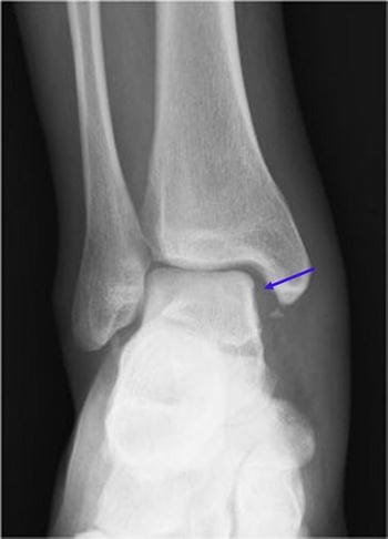
Figure 2: Widening of the ankle mortise
Oftentimes in the ED, you will be clued into a possible Maisonneuve fracture with the presence of the proximal fibular fracture. This is key to identify in your physical exam. A complete physical exam to evaluate ankle injuries should include evaluation of the proximal fibula. When you see this, your attention should immediately turn to the ankle x-ray, looking for a possible medial malleolus fracture. If no fracture exists, a stress-view XR of the ankle mortise should be obtained to identify possible widening (Figure 2). This can be done by having the patient stand, for weight bearing films. If a patient cannot stand, you may manipulate the ankle, adding gentle pressure as you place the ankle in dorsiflexion and internal rotation. If you see an increase in the medial clear space, this likely indicates injury to the tibiofibular syndesmosis, and thus you have identified a probable Maisonneuve fracture.
The injury pattern involved with a Maisonneuve fracture creates an unstable ankle, as the syndesmosis is violated. Thus, treatment of Maisonneuve fractures often involves operative repair. (5) In the ED, the affected leg should be placed a posterior long leg splint, with efforts to reduce the syndesmosis to anatomical alignment. The patient should be made non-weight bearing and given crutches. An orthopedist should be contacted to ensure urgent follow up for definitive repair.
Case Wrap Up
Now that you’ve identified that your patient has a Maisonneuve fracture, he should be placed in a long leg splint. He should be counseled that this will likely require operative management and instructed not to bear weight on his right ankle. An orthopedics consult should be placed to ensure urgent follow up as this is an unstable injury. Patient would be appropriate for discharge once follow up is arranged.
References
1. Silverman ARC, Broome JN, Jarvis RC 3rd, Plassche GC, Kirschenbaum IH. Using Emergency Department Data to Inform Specialty Strategy: Analyzing the Distribution of 13,777 Consecutive Immediate Orthopaedic Consults in an Urban Community Emergency Department. J Am Acad Orthop Surg Glob Res Rev. 2020 Feb 12;4(2):e20.00005. doi: 10.5435/JAAOSGlobal-D-20-00005. PMID: 32440626; PMCID: PMC7209810.
2. Zhao B, Li N, Cao HB, Wang GX, He JQ. Rare pattern of Maisonneuve fracture: A case report. World J Clin Cases. 2022 May 16;10(14):4684-4690. doi: 10.12998/wjcc.v10.i14.4684. PMID: 35663082; PMCID: PMC9125267. (Maisonneuve fx’s account for approx. 7% of all ankle fx’s)
3. He JQ, Ma XL, Xin JY, Cao HB, Li N, Sun ZH, Wang GX, Fu X, Zhao B, Hu FK. Pathoanatomy and Injury Mechanism of Typical Maisonneuve Fracture. Orthop Surg. 2020 Dec;12(6):1644-1651. doi: 10.1111/os.12733. Epub 2020 Sep 7. PMID: 32896104; PMCID: PMC7767678.
4. Dr. Sachin Kale, Dr. Pratik Tank, Dr. Rahul Ghodke, Dr. Pankaj Singh, Dr. Abhiraj Patel. Functional outcome of bimalleolar ankle fractures treated with fibular plate for lateral malleolus and C.C. screws for medial malleolus. Int J Orthop Sci 2020;6(1):1126-1132.
5. Stufkens SA, van den Bekerom MP, Doornberg JN, van Dijk CN, Kloen P. Evidence-based treatment of maisonneuve fractures. J Foot Ankle Surg. 2011 Jan-Feb;50(1):62-7. doi: 10.1053/j.jfas.2010.08.017. PMID: 21172642.
Authorship
Written by - Anthony Martella, MD, PGY-4, University of Cincinnati Department of Emergency Medicine
Peer Review by
Bret Betz, MD, Assistant Professor, University of Cincinnati Department of Emergency Medicine
Daniel Gawron, MD, Sports Medicine Fellow, Assistant Professor, University of Cincinnati Department of Emergency Medicine
Editing and Posting - Jeffery Hill, MD MEd, Associate Professor, University of Cincinnati Department of Emergency Medicine
Cite As
Martella, A. Hill, J. Gawron, D, Betz, B. Twists and Turns: Identifying Maisonneuve Fractures in the ED. www.tamingthesru.com/blog/maisonneuve-fractures. TamingtheSRU. 9/25/23


