Air Care Series: Critical Care Transport Medicine (CCTM) Ultrasonography: Past, Present, and Future
/As the saying goes, a picture is worth a thousand words—but a picture might be worth even more on a helicopter where the ability to hear and see is limited. Given the challenges of the operating environment, clinicians have been working to implement POCUS in critical care transport medicine (CCTM) for more than two decades.[1] At UC Health Air Care, flight physicians have had access to ultrasound on CCTM missions for over a year, and we wanted to share cases where ultrasonography has proved its mettle.
We are believers that this is the future: not just for flight physicians, but for all HEMS/CCTM colleagues— see this recent report of Taming the SRU contributor and University of Cincinnati Emergency Medicine alumnus Andrew Latimer, MD and colleagues describing an initial ultrasound training curriculum for flight nurses.[2]
Evidence from a Danish registry suggests providers use ultrasound on more than 20% of CCTM air transports.[3] While various protocols have been described for emergency ultrasound use in the CCTM environment, the typical areas of interest have been to evaluate for intra-abdominal hemoperitoneum, hemopericardium/pericardial effusion, intra-thoracic traumatic injuries including pneumothorax, and confirmation of endotracheal intubation.[4,5] While much focus has been on trauma patients’ evaluation, publications from the University of Pittsburgh have examined the utility for medical patients—including dyspneic patients and out-of-hospital cardiac arrest (OHCA).[6,7] With that background, what kinds of things have the early-adopter flight docs at AirCare seen so far?
Case #1:
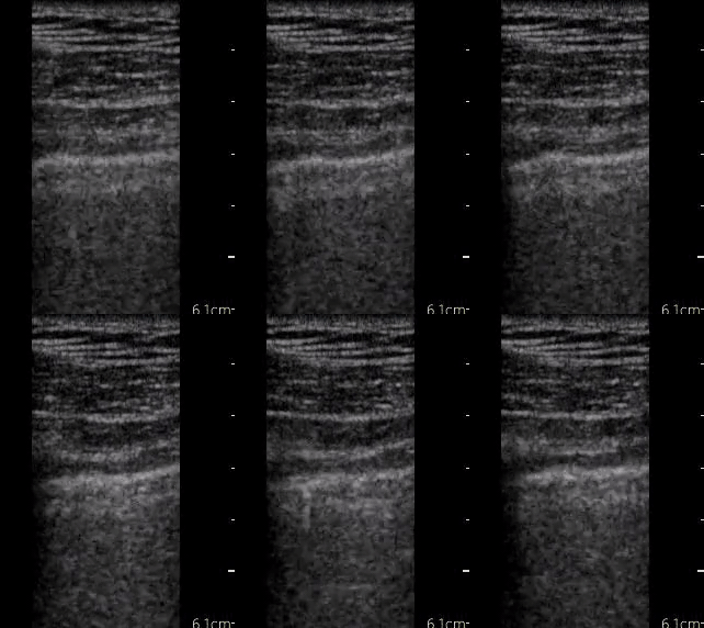
A middle-aged man fell from a roof at a distance estimated by scene EMS to be ~30 feet. The patient complained of left-sided chest pain, but initial vitals including RR and O2 sat were reassuring. During rotor-wing transport, the patient’s oxygenation saturation decreased from 98% to 91% on room air.
The flight physician obtained the following clip on the affected side
+ What do you see?
Yep, a normal thoracic ultrasound evaluation with lung sliding seen. A mid-flight needle or open thoracostomy was avoided
Case #2:
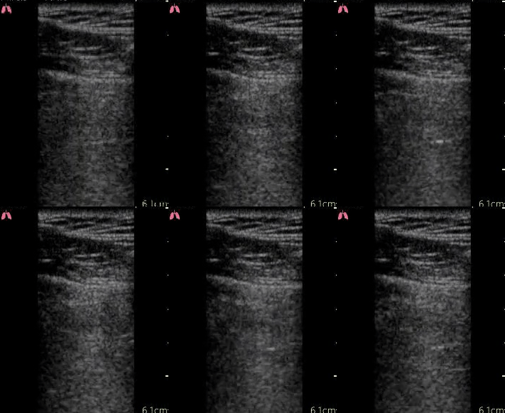
An elderly man was struck in the head and chest by piece of farming equipment while repairing it. The patient had left-sided head, chest, and shoulder pain, which were all severe. The patient was hypoxemic for responding EMS, and was placed on supplemental oxygen by nasal cannula, which was continued by the CCTM crew.
In-flight, the CCTM crew obtained the following images
+ What do you see?
That is a left sided pneumothorax. The patient remained hemodynamically stable and free from worsening hypoxemia on the nasal cannula. CT scan confirmed left 1-3 rib fractures with a pneumothorax. A chest tube was placed under sterile conditions in the Emergency Department
Case #3:
An otherwise healthy young male reported sudden onset chest pain and dyspnea on arrival to a free-standing Emergency Department. A CT scan showed no pulmonary embolism but did find what was thought by the referring ED physician to be a ruptured spleen for which transfer had been arranged. The patient strictly denied any trauma, and final radiology interpretation was still pending.
In concert with the referring doctor, the crew obtained a single clip of the RUQ, what do you think?
+ What do you see?
Yep, that’s a positive FAST. The etiology of the spleen rupture remained uncertain, but the referring doctor (and the flight crew) now have evidence consistent with a potentially spontaneous splenic rupture with hemoperitoneum.
In addition to diagnostic information, there is significant opportunity to facilitate procedural tasks with ultrasound. For patients with difficult vascular access, we have developed a protocol to allow placement of a peripheral intravenous catheter in the internal jugular vein to allow appropriate, expeditious, and reliable access prior to flight or during transport.
We know ultrasound can and will help us deliver optimal critical care for our patients – not just helping us find the correct thoracic and pericardial spaces that do (and do not) need intervention, but also helping identify the presence of internal hemorrhage so that a lowering blood pressure merits transfusion. Even before a patient arrives, we can mobilize resources in advance at the receiving hospital by identifying Trauma STAT criteria (e.g., a positive FAST exam)…and many more applications we are excited to be able to offer our patients!
With all our enthusiasm for Ultrasound in the HEMS setting, we’re cognizant that increased on-scene time in unstable trauma patients is potentially detrimental to outcome.[8] But our early experiences have convinced us that the future is bright for US in HEMS… (and if it’s too bright, we can always turn down the gain!).
AUTHORED BY Bennet Lane, MD MS & Adam GotTula, MD
Dr. Lane is a recent graduate of University of Cincinnati Emergency Medicine Residency Program. He is now an Assistant Professor of Emergency Medicine at the University of Cincinnati
Dr. Gottula is a recent graduate of University of Cincinnati Emergency Medicine Residency Program. He is now at the University of Michigan completing a fellowship in Critical Care.
FACULTY EDITOR Shaun Harty, MD
Dr. Harty just completed an ultrasound fellowship at the University of Cincinnati
References
1. Melanson SW, McCarthy J, Stromski CJ, Kostenbader J, Heller M. Aeromedical Trauma Sonography by Flight Crews With a Miniature Ultrasound Unit. Prehosp Emerg Care. 2001;5(4):399-402. doi:10.1080/10903120190939607
2. Mason R, Latimer A, Vrablik M, Utarnachitt R. Teaching Flight Nurses Ultrasonographic Evaluation of Esophageal Intubation and Pneumothorax. Air Med J. 2019;38(3):195-197. doi:10.1016/j.amj.2018.11.007
3. Alstrup K, Møller TP, Knudsen L, et al. Characteristics of patients treated by the Danish Helicopter Emergency Medical Service from 2014-2018: a nationwide population-based study. Scand J Trauma Resusc Emerg Med. 2019;27(1):102. doi:10.1186/s13049-019-0672-9
4. Chin EJ, Chan CH, Mortazavi R, et al. A Pilot Study Examining the Viability of a Prehospital Assessment with UltraSound for Emergencies (PAUSE) Protocol. J Emerg Med. 2013;44(1):142-149. doi:10.1016/j.jemermed.2012.02.032
5. Bhat S, Johnson D, Pierog J, Zaia B, Williams S, Gharahbaghian L. Prehospital Evaluation of Effusion, Pneumothorax, & Standstill (PEEPS): Point-of-care Ultrasound in Emergency Medical Services. West J Emerg Med. 2015;16(4):503-509. doi:10.5811/westjem.2015.5.25414
6. Becker TK, Martin-Gill C, Callaway CW, Guyette FX, Schott C. Feasibility of Paramedic Performed Prehospital Lung Ultrasound in Medical Patients with Respiratory Distress. Prehosp Emerg Care. 2018;22(2):175-179. doi:10.1080/10903127.2017.1358783
7. Fitzgibbon JB, Lovallo E, Escajeda J, Radomski MA, Martin-Gill C. Feasibility of Out-of-Hospital Cardiac Arrest Ultrasound by EMS Physicians. Prehosp Emerg Care. 2019;23(3):297-303. doi:10.1080/10903127.2018.1518505
8. Harmsen AMK, Giannakopoulos GF, Moerbeek PR, Jansma EP, Bonjer HJ, Bloemers FW. The influence of prehospital time on trauma patients outcome: A systematic review. Injury. 2015;46(4):602-609. doi:10.1016/j.injury.2015.01.008


