Diagnostics: Brain MRI

Diagnostics: Brain MRI

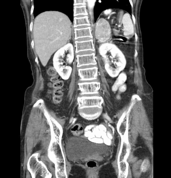
If you have ever nodded along as someone described an MRI series weighting, you are not alone. As this modality becomes more accessible to the Emergency Physician, it is incumbent on us to familiarize ourselves with the critical aspects and critical diagnoses of MRI and how to speak the language. Dr. Zalesky takes us on a tour of the physics, indications and low-down of MRI from the ED.

Read More

X-ray Vision - Shoulders and Elbows

X-ray Vision - Shoulders and Elbows

Upper extremity trauma and pain related complaints are frequently encountered in the Emergency Department. In this post, we cover the basics of the anatomy of the shoulder and elbow joint, the radiographic studies frequently performed in the evaluation of shoulder/elbow injuries, and cover some commonly encountered injuries of these joints.

Read More

Back to Basics: Pelvic XRays

Back to Basics: Pelvic XRays

Pelvic Xrays are a key component of trauma, fractures and dislocations seen every day in the ED, but when is the last time you went back over the anatomy and radiographic tips and tricks of the pelvic radiograph? Join Dr. Mand's thorough break down of this commonly used ED diagnostic - the Pelvic XR.

Read More

Soft Tissue Neck Radiographs - Are They Useful?

Soft Tissue Neck Radiographs - Are They Useful?

In March 2015, Dr. Renne did a Grand Rounds lecture on soft tissue neck radiographs, which offers a great review of normal anatomy and systematic approach to reading films (“CHESS”). Yet, in my small, informal (not scientific at all) poll of some of our residents, none had ever ordered a soft tissue neck film. Are soft tissue neck radiographs useful? You be the judge. 

Read More

Interpreting Chest X-rays

Interpreting Chest X-rays

There isn't a day that goes by in the ED that a patient does not get a chest x-ray.  Whether the indication is chest pain, shortness of breath, cough, or line placement or intubation, interpreting chest radiographs is a critical, necessary skill for anyone working in the Emergency Department.  Here you will find a brief video explaining how to interpret CXRs and 6 practice cases.

Read More

The Approach to the Foot X-ray

The Approach to the Foot X-ray

Any way you slice it, foot x-rays are a pain to read.  Complicated by a number of overlapping bones, joints, the presence of multiple sesamoid bones, and multiple radiographic views, it's easy to get lost in the weeds trying to sort out normal variant from pathology.  Take a look at this short Blendspace module by PGY-1 Lauren Titone, MD and get a better understanding of the normal anatomy and a systematic approach to reading foot x-rays.

Read More

Grand Rounds Recap - 11/13/14

Grand Rounds Recap - 11/13/14

SBIRT (Screening, Brief Intervention, Referral, & Treatment) for Substance Abuse

Why should we care?

  • Prevalence of this disease is impressive with greater than 33,000 deaths attributed to alcohol in 2012 alone (287,000 MVC's in Ohio alone attributable to alcohol)
  • Medical problems attributable to alcohol use costs the US $100,000,000,000 annually (from health care bills to lost productivity)!
  • Approximately 33% of inpatient admissions in a country hospital population were attributable to alcohol
  • One in five Americans can be defined as at risk drinkers
Read More

Interpretation of Ankle Radiographs

Interpretation of Ankle Radiographs

A 43 year old female presents to the ED after "rolling" her ankle while gardening.  She states that she was stepping down on a shovel when her ankle rolled.  <She describes to you and inversion type injury.>  Being a diligent, studious physician, you quickly run through the Ottawa Ankle Rules while you obtain the remainder of you history and physical.  She was unable to bear weight immediately after the accident and is, likewise, unable to do so here in the ED.  She has no pain with palpation over the medial malleolus but does have significant pain and tenderness with palpation of the lateral malleolus.  You quickly decide that this patient will need ankle radiographs to further investigate the possibility of fracture.  

But, what views should you order? And, once you get the films back, how do you interpret them.  Check out the excellent video embedded below, made by Dr. Claire O'Brien, PGY-1 in the University of Cincinnati Dept. of Emergency Medicine Residency Training program, to find out!

Read More