Ultrasound of the Month: Lipohemarthrosis
/THE CASE
A female in her 70’s with a past medical history of atrial fibrillation on apixaban, hypertension, hyperlipidemia, and a previous total right knee arthroplasty presented to the emergency department after a fall. She experienced a ground level fall onto a curb earlier in the day and has been unable to bear weight since then. On arrival at the emergency department, she reports pain in her right lower extremity including her knee, lower leg, and foot.
On exam, she was noted to have swelling in the knee and ecchymosis in the knee and lower leg. She was able to range the knee but with significant pain. Her sensation was intact to light touch. On examination of her pulses, her right dorsalis pedis was diminished. Plain radiographs of the knee were obtained that demonstrated a joint effusion tracking along the patella but no bony abnormality. Given her asymmetric pulses, a computed tomography of the aorta (CTA) with runoff down to both lower extremities was obtained. Due to the concern for an effusion on exam, an ultrasound of the right knee was performed and the images are below.
Image 2: : A longitudinal view of the right knee demonstrating lipohemarthosis with layering of fat, serum and RBCs
Image 1: A transverse view of the right knee demonstrating lipohemarthrosis with layering of fat, serum and RBCs
PATHOLOGY OF LIPOHEMARTHROSIS
Traumatic injuries to the knees are incredibly common in the emergency department, accounting for over half a million visits annually [1]. Initially, most providers evaluate the patient with plain film radiography, which may miss occult fractures. In response to trauma, the joint often accumulates fluid as a reactive response, creating a joint effusion. Not all effusions, however, are simple fluid collections. If there is an intra-articular fracture of the bone, such as a tibial plateau fracture, blood in addition to fat from the bone marrow may seep into the joint space, creating a lipohemarthrosis. In this setting, ultrasound may be used to evaluate the joint effusion for fat globules and blood in the joint, which is a surrogate marker for fracture [2].
On ultrasound, lipohemarthrosis appears as a joint effusion with a distinct fluid-fluid level that is created from the separation of fat and blood components. Fat, less dense than blood and synovial fluid, makes up the most superficial layer, which appears as a hyperechoic band floating atop of the effusion. Beneath this lies a more hypoechoic layer, representing the serous component of blood and synovial fluid. Two to three hours following injury, the blood begins to separate into its constituent parts; the serum and erythrocytes [1,2,3,4]. The red blood cells may then settle to form a third dependent layer, which appears as a mildly hyperechoic strip deep to the anechoic fluid. The layered appearance is demonstrated below using images from our patient.
Image 3: Longitudinal view of the knee demonstrating the layers of lipohemarthrosis (fat, serum and layered RBCs superior to the anterior cortex of the femur)
Additionally, ultrasound may visualize the fracture directly by showing a disruption in the smoothness of the bony cortex. Ligamentous injuries may be similarly seen with disruption of the fibers of the ligament.
IMAGING WITH ULTRASOUND
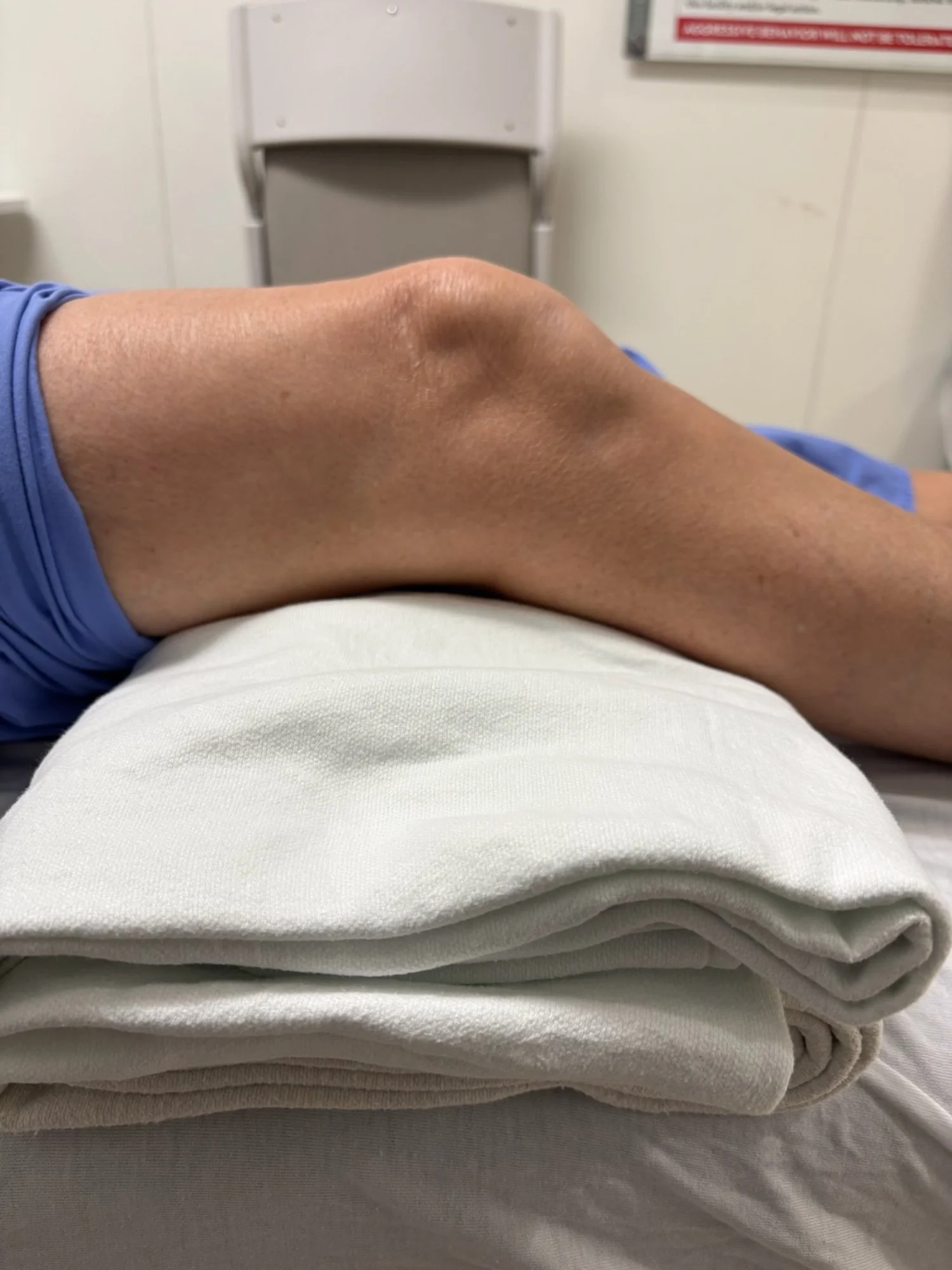
When performing joint ultrasound, placing the knee in 30 degrees of flexion provides the best orientation for imaging joint fluid [2]. This can be achieved by placing a pillow or blanket roll under the knee (image 4). When scanning transversely, one should interrogate the medial and lateral joint lines, especially when evaluating for an arthrocentesis pocket. Then, the probe can be rotated into the longitudinal orientation. In this view, starting from the base of the patella and moving cranially, one can evaluate the tendon, patellar cortex, femoral cortex, and the suprapatellar pouch [2].
Ultrasound is not routinely used to image the knee joint when searching for fracture. However, it is used to evaluate the quality of a joint effusion and to assess for an acceptable pocket for arthrocentesis. When evaluating joint effusions, most will be anechoic as the synovial fluid should have minimal particulates present. In the setting of blood or infection, particulate matter may be seen floating inside the effusion [2].
If one notes concern for lipohemarthrosis on ultrasound, this should prompt the consideration of additional evaluation for an occult fracture, particularly of the tibial plateau. The standard imaging modality is a non-contrast CT scan. Tibial plateau fractures are unstable and carry a high rate of compartment syndrome. In many cases, patients may be admitted for compartment checks overnight during the acute swelling phase post injury.
CASE RESOLUTION
Despite negative radiography, given the patient’s abnormal exam and ultrasound findings, the orthopedic team was consulted and a non-contrasted CT of the knee was ordered in addition to the CTA. The CTA demonstrated no vascular injury but the non-contrast CT did demonstrate a nondisplaced fracture through the distal femoral metaphysis with a large associated lipohemarthrosis. She was admitted to orthopedic surgery and was managed nonoperatively. After being assessed by PT/OT, she was discharged to a SNF for further therapy.
AUTHORED BY Kelly Tillotson, MD
Dr. Tillotson is an EMS Fellowship trained graduate of Emergency Medicine at the University of Cincinnati
PEER REVIEW By Martina Diaz McDermott, MD
Dr. McDermott is an Ultrasound Fellowship trained graduate of Emergency Medicine at the University of Cincinnati
EDITING AND LAYOUT BY Olivia Gobble, MD
Dr. Gobble is a current Ultrasound fellow at the University of Cincinnati and recent graduate of Emergency Medicine at the University of Cincinnati
REFERENCES
Elisa M. Aponte, Joseph I. Novik, Identification of Lipohemarthrosis With Point-of-Care Emergency Ultrasonography: Case Report and Brief Literature Review, The Journal of Emergency Medicine, Volume 44, Issue 2, 2013, Pages 453-456, ISSN 0736-4679,
Rippey J. Ultrasound for knee effusion: lipohaemarthrosis and tibial plateau fracture. Australas J Ultrasound Med. 2014 Nov;17(4):159-166. doi: 10.1002/j.2205-0140.2014.tb00239.x. Epub 2015 Dec 31. PMID: 28191232; PMCID: PMC5024930.
Costa DN, Cavalcanti CF, Sernik RA. Sonographic and CT findings in lipohemarthrosis. AJR Am J Roentgenol. 2007;188(4):W389. doi:10.2214/AJR.06.0975
Gaillard F, Yu Jin T, Murphy A, et al. Lipohemarthrosis. Reference article, Radiopaedia.org (Accessed on 27 Nov 2023) https://doi.org/10.53347/rID-1358
Academic Life in EM: Use of Point-of-care Ultrasound in Tibial Plateau Fracutres https://www.aliem.com/ultrasound-tibial-plateau-case/


