Please see the Annals of B Pod article on central line placement in order to review indications for this procedure and how to select an appropriate catheter.
Selecting an Insertion Site
Subclavian Surface Anatomy
Read the following posts on the relevant anatomy for femoral and subclavian access:
Anatomy of a Procedure: Subclavian Access
Anatomy of a Femoral Venous Access
Preparation
Assess the anatomy of the selected insertion site and evaluate it using ultrasound prior to starting procedure.
Identify necessary PPE- wash hands, wear cap, mask, sterile gown, and sterile gloves. Use sterile probe cover for the ultrasound.
Clean the insertion site with chlorhexidine.
Place a full drape on the patient.
Maintain sterile field throughout the procedure.
Prepare equipment including flushing lines and placing caps on the ports.
Vessel Cannulation
Identify the needle insertion site that will allow cannulation of the desired vein.
Anesthetize the area over the insertion site.
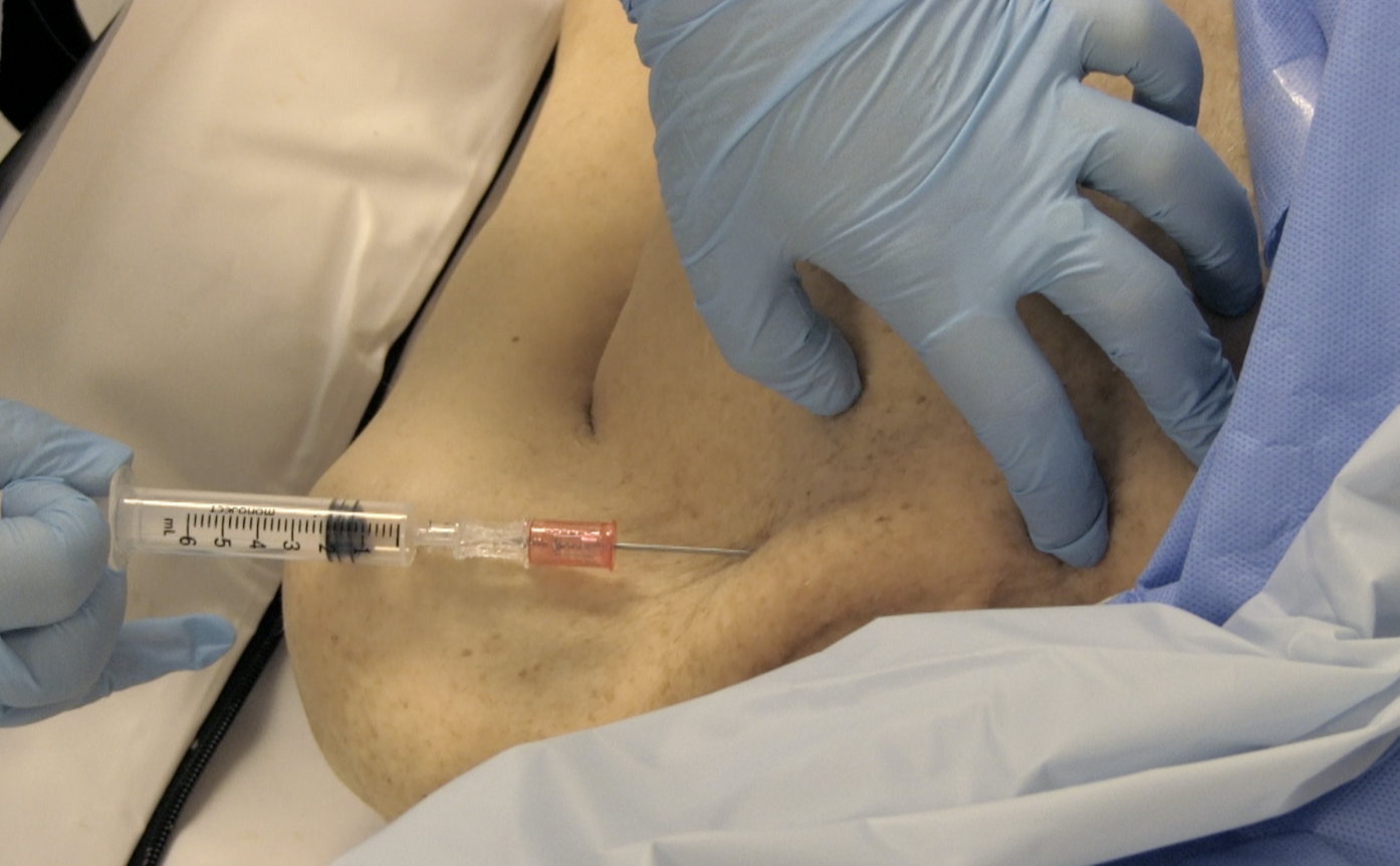
Insert and advance the needle while continuously aspirating.
Follow the needle tip into the vessel using ultrasound guidance. Needle visualization is a critical micro skill in the performance of any ultrasound-guided procedure. Please watch this video from Dr. Stolz on how to improve your needle visualization.
Successfully cannulate the vessel and ensure free return of dark red, non-pulsatile blood.
Stabilize the needle while disconnecting the syringe.

Threading The Wire
Insert guidewire through the needle to a depth of 20 cm.
Remove needle while maintaining control of the guidewire.
Confirm guidewire is in the vessel using ultrasound.


Dilating the Vessel and Subcutaneous Tissues
Make small skin incision adjacent to the guidewire.
Advance dilator over the guidewire and into skin with a twisting motion while maintaining control of the guidewire.
Ensure the guidewire moves freely within the dilator, then remove dilator.


Advance the Catheter over the Guidewire
Advance catheter over the guidewire and into the vessel to the appropriate depth for the selected insertion site.
Remove guidewire and keep the brown port covered to prevent air entry until a cap can be placed on the brown port.
Aspirate and flush each lumen.


Securing the Line
Suture the line in place at the blue/white clips and the triangular holder.
Add biopatch and place a sterile dressing.
Obtain CXR following the procedure.
Other Links from Around the web
NEJM Videos in Clinical Medicine Series
EMCrit on Central Lines - https://emcrit.org/central-lines/
5MinSono
References
Kim, Won Young et al. (2012) Optimal insertion depth of central venous catheters—Is a formula required? A prospective cohort study. Injury , Vol. 43(1), 38-41.
Comments, Suggestions, Feedback? Send us a Message!


