Grand Rounds Recap 9.7.2016
/ClinicoPathologic Case Conference: Dr. Shaw versus Dr. Doerning
The Case
A young female with bipolar disorder, psychosis, anemia, GERD who presents with rib pain. The pain is constant but worsens for about 60 seconds at a time at random. Pain is present over the 5th right rib in the mid-axillary line on palpation.
CXR - normal
EKG - shows lead III, V3-V6 show T wave inversions concerning for right heart strain
PERC
- if negative: <2% chance of PE if pre-test probability <15%
- considered to be the point of equipoise for which risk of PE/harm of CTPA is balanced
Well’s Score of 0: 1.3% chance of PE conditional on low pre-test probability
A retrospective analysis of the combined use of PERC rule and Wells score to exclude PE in the ED
21% of patients in the study with a Wells < 2 and negative PERC: 2% had an elevated d-dimer and subsequently 2 CTPAs showed a sub-segmental PE (0.6%). The question is does this subsegmental PE matter?
In this case the providers felt that PE was at least as likely any other diagnosis (particularly given the EKG findings) which put the patient into the moderate risk Wells category, for which PERC would not be appropriate.
Diagnostic test of choice: d-dimer which came back slightly elevated.
CTPA showed segmental PE, subsegmental PE, pulmonary infarction, and evidence of retrograde blood flow into hepatic veins suggestive of right heart strain.
Findings on EKG in PE:
- Tachycardia
- Sinus
- Atrial tachycardia: fib and flutter
- Right Heart Strain
- RBBB
- Right axis deviation
- S1, Q3, T3
- Inverted T waves can occur together in the inferior and antero-medial precordial leads (V1-V4) in R heart strain - which is less likely with ACS
Evaluation of PE in pregnant patients:
- D-dimer is less useful as a rule-out in: by the time the patient is in the second trimester only 22% of patients will have a negative d-dimer, by the third trimester almost no pregnant patients have a negative d-dimer. Maybe there will be a pregnancy adjusted d-dimer someday soon, however it’s not yet validated.
- Options for diagnosis: CTPA and VQ Scan
- Fetal radiation is slightly higher in VQ scan versus CTPA
- Breast cancer risk is 150 times higher for CTPA versus VQ scan
- If adequate and normal CXR - VQ scan is more likely to be diagnostic than CTPA in pregnant patient
- OBs prefer CTPA because fetal radiation studies are mostly theoretical and there is concern that the radioactive tracer used in VQ would be more harmful to the fetus when it collects in the bladder. This is ultimately poorly studied but if you are to get a VQ put in a foley which also should reduce the fetal dose
PE in the anti-coagulated patient:
- Compliant patients: may be treatment failure. They need to be switched to a different agent
- Non-compliant patients: restart their anti-coagulation
Hand and Wrist X-rays with Dr. Michael Spigner
How to read hand and wrist films.
Lunate Dislocation:
- Lunate disarticulates from both the radius and capitate. "Tipped teacup sign"
- Acute carpal tunnel syndrome is a possible complication
- Often requires surgical repair
Peri-lunate Dislocation:
- Lunoradial articulation is intact, but articulation with the capitate and remaining carpal bones is lost
- 60% associated with scaphoid fracture
- Median nerve may also be involved
Scaphoid Fractures:
- If films negative: splint and re-examination in 10-14 days and MRI are considered equivalently appropriate therapies
- Can consider advanced imaging depending on resource availability in a patient who returns in 10-14 days with continued clinical suspicion but negative films (MRI is slightly better than CT)
Carpometacarpal Dislocation:
- Commonly associated with base of metacarpal fracture
- Often hard to reduce/maintain reduction: 50% require ORIF
EBM Lecture: Sexually Transmitted Infections with Drs. Continenza and Randolph
History Taking: Ask about the 5 Ps
- Partners
- Practices
- Prevention of pregnancy
- Protection from STIs
- Past history of STIs
Use a recommended treatment regimen: CDC Guidelines and App
Always perform a pelvic exam if any concern for PID
- If asymmetric tenderness on adnexal exam or other concern for TOA or for non-gynecologic surgical emergency, consider additional imaging with CT or Ultrasound. CT is slightly more sensitive than ultrasound for detecting TOA
- Minimum Exam Criteria for PID (at least one of the following)
- Cervical motion tenderness
- Uterine tenderness
- Adnexal tenderness
- Fever and elevated WBC count are not required but can add specificity
- "Chandelier sign" is less common as the incidence of PID caused by gonococcal infection has decreased
- CDC recommends empiric treatment of all women with risk factors, lower abdominal pain, at least one of the minimum criteria and no more likely explanation for symptoms
STIs can occur in the rectum and oropharynx so don't forget to ask about sexual practices and perform the appropriate exams
In transgender patients, focus on their anatomy and practices. Gender association is not the same as sexual orientation
Special Populations:
- MSM, MSMW are high risk for HIV, syphilis, resistant bacterial, pharyngeal and rectal pathology (http://www.lgbthealtheducation.org)
- Sex-trafficked patients: at high risk for loss to follow up, recurrent infections, abuse
- Adolescents: you do not need parental consent to test or treat
R4 Case Follow-Up with Dr. Thomas
Neonatal Resuscitation: Key Points
First: Assess tone, cry, color. Stimulate, warm, and suction (10 seconds)
Airway
- Put baby's head in a "sniffing position" and bulb-suction mouth/nose
- PPV with FiO2 21-30% if meconium present, suction with bulb syringe NOT tracheal suctioning
- Intubation only after failing PPV
Breathing
- PPV first for apnea, gasping, or pulse < 100 bpm
- Ventilate at a rate of 40-60 breaths per minute
- Expect lower oxygen saturations in the first 10 minutes of life
Circulation
- Start chest compressions (two thumb wrap-around method) only after > 30 seconds of PPV
- Ideally compressions start with advanced airway in place
- Ratio: 3 compressions to 1 breath
Drugs
- Epinephrine if HR < 60 after 60 seconds of compressions
- Fluids or blood if suspected hypovolemia
- Naloxone - not recommended
- Glucose - not recommended
Pulse < 60 bpm: Resuscitation
- Begin compressions
- 3:1 Ratio
- 120 events per minute
- Increase FiO2 to 100%
- Vascular access (consider umbilical line)
- Pulse check every 60 seconds
- Initiate pharmacology after the first 60 seconds
Pediatric Hematologic Emergencies with Dr. Matthew Scanlon
Hyperleukocytosis and Leukostasis
- Definition: WBC > 100,000
- Common in AML, ALL - nearly always as the result of a malignancy
- Blood becomes viscous causing multiple complications
- Stroke from stasis in cerebral blood vessels
- Bone pain from marrow expansion
- Pulmonary infarcts and infiltrates
- Limb ischemia, bowel infarction, priapism
- Fevers are common
- Suppression of other cell lines can cause anemia, thrombocytopenia, DIC
- Spontaneous tumor lysis syndrome can occur due to rapid cell turnover
- Management:
- Admission and emergent heme/onc consultation for rapid chemo induction (mortality is 20-40% in one week if not treated)
- CBC, PT/INR, Fibrinogen
- Prophylactic platelet transfusion recommended to 20-30,000
- Judicious fluid resuscitation
- Stroke: tPA not indicated - the stroke is causes by stasis of blood flow not thromboembolism
Acute Chest Syndrome
- Vaso-occlusion within the pulmonary vasculature leads to ischemia and endothelial injury
- Precipitated by: unknown etiology (46%), infections (29%), pulmonary infarction (16%), fat embolism secondary to avascular necrosis
- Other risk factors: asthmatic disease, chronic hypoxemia, post-op
- Diagnostic criteria: New pulmonary infiltrate on CXR and at least one of the following
- Chest pain
- Temp > 38.5 C
- Respiratory distress
- Hypoxemia
- Management: fluids, antibiotics (3rd gen cephalosporin + macrolide), respiratory support, pain control
- Transfusion may be indicated if profoundly anemic
Aplastic Crisis
- Complication in Sickle Cell Anemia
- Most common cause is suppression of erythropoiesis due to Parvovirus B19
- Presents with pallor, weakness, lethargy
- CBC shows > 30% in hemoglobin from baseline and critically low or absent reticulocytes
- Management: simple transfusion
Hemophilia
- In the ED it's important to recognize a first bleed in a patient who does not carry the diagnosis
- Cephalohematoma at birth can be a clue
- Hemarthrosis
- Forehead hematomas
- Remember: There are NO trivial head injuries in patients with hemophilia. Have a low threshold for imaging and repletion
- Management: Replete factor to greater than 50% activity (consult pharmacy or use patient's own supply), steroids for hemophilic arthropathy
Sepsis and qSOFA with Dr. Walsh
Goal of the guidlines: Differentiate sepsis from uncomplicated infection, update definitions of sepsis and septic shock
Sepsis is a syndrome without a validated, goal standard test
SIRS criteria are not specific to sepsis and not all inflammation is bad, whereas all organ dysfunction is bad
Sepsis by definition involves organ dysfunction secondary to dysregulated physiologic response to infection and is always life threatening. The new guidelines throw out the classification of severe sepsis, all sepsis is severe
SOFA Score
- Measures organ dysfunction and outperforms SIRS in predicting hospital mortality.
- Organ dysfunction defined as increase in SOFA score by 2 over baseline.
- General hospital population with SOFA score >= 2, Mortality = 10%
qSOFA: quick SOFA score
- Respiratory rate > 22/min
- Altered mentation
- Systolic BP < 100 mmHg
Septic Shock:
- Persistent hypotension requiring vasopressors to maintain map > = 65
- Serum lactate level > 2
- Define only after "adequate" fluid resuscitation (which is not defined).
Limitations:
- SOFA requires extensive laboratory testing (qSOFA does not)
- These are not standalone and should not be treated as screening tests
Fever in the Returning Traveler with Dr. Wright
Most common presentations in ill travelers: GI distress, fever, and rash
Diarrhea:
- Characteristics/testing of the diarrhea can be helpful
- Blood: inflammation
- WBCs: bacteria
- Ova and parasites
- Gram stain: Campylobacter
- Culture: Salmonella, Shigella, Campylobacter
- Special culture: Yersinia, Vibrio
- Trichrome stain: microsporidia
- Safranin: Cyclosporia
- ELISA: Giardia, crypto, EC O157:H7, Entamoeba, C. diff
- Management: supportive
- Hydration
- Empiric antibiotics
- Africa or Latin America: Fluoroquinolones
- Southeast Asia: Macrolide
- Anti-motility agents: controversial and definitely not for < 3 years of age
Vibrio Cholera Outbreak in Haiti:
- Characteristics: Watery ("Rice water") diarrhea, 3-5 day incubation period, vomiting and muscle cramps
- Introduced by UN peacekeepers, 700,000 affected, still ongoing
Fever:
- Marker of potentially life threatening illness in a returning traveler
- SE Asia, Carribean, Latin America - Dengue
- Africa - Malaria
- Spread by anopheles mosquito
- More common, more deadly: falciparum and malariae
- Less common: vivax, ovale, knowlesi
- Incubation 3-5 days (falciparum is faster) can take months for the others (vivax, ovale)
- Drugs:
- Malarone is the currently recommended prophylaxis and therapy
- Coartem
Work up for fevers in returning travelers:
- All should get: CBC, renal, hepatic, UA, CXR
- Some should also get: thick/thin smear and rapid diagnostic testing (if exposed to malaria)
- Long duration travelers: test for TB using quantiferon gold
- Serologies: dengue, rickettsia, fungus, lyme, brucella, arbovirus, leptospirosis, filariasis, schistosomiasis
Rashes:
Measles
- Highly infectious 90% of those exposed will develop disease
- Incubates for 6-21 days, prodrome is 2-4 days
- Somewhat unique in that it comes with a cough
- Also conjunctival injection and classic rash
- Complications include encephalitis
- Reportable disease
Dengue
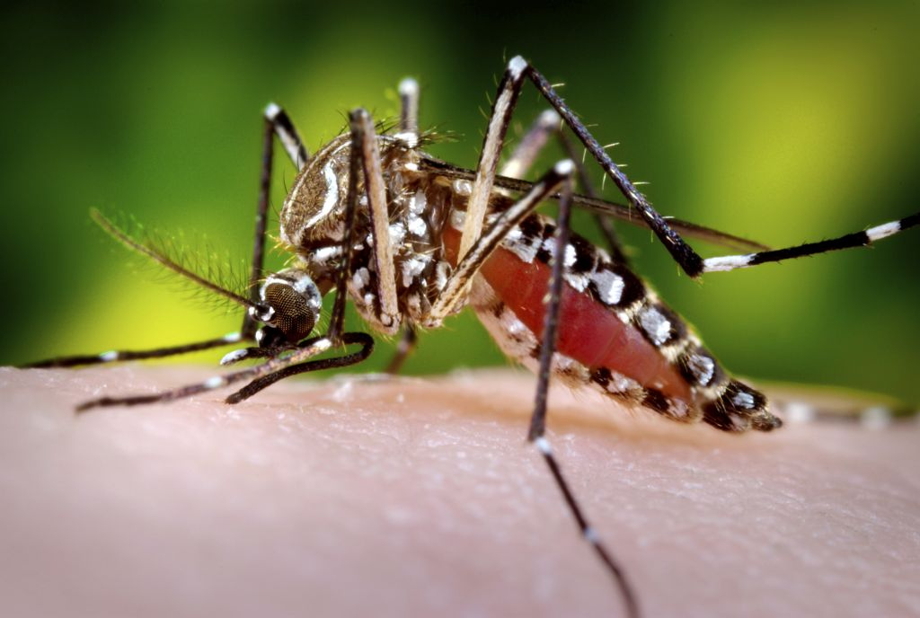
- rash with “islands” of spared skin, spread by Aedes mosquito
- breakbone fever, headache, eyepain, mild bleeding, and leukopenia
- "Tourniquet Test": petechiae may develop under tourniquet
- may want to get INR and platelet count before deciding about LP
Zika:
Complications:
- Microcephaly
- Guillain-Barre syndrome
Pregnancy advise after travel to an endemic area or exposure to Zika:
- Wait 6 weeks if asymptomatic
- Symptomatic Women: wait 8 weeks after travel for pregnancy (for symptoms to develop)
- Symptomatic Men: should wait 6 months before attempting to conceive
- Condoms to prevent transmission for the same period of time
- Zika Algorithm
Schistosomiasis:
- Swimmer’s Itch - the fluke entering your skin after touching/swimming in infested water
- Acute schistosomiasis syndrome: katayama fever is a hypersensitivity reaction to antigens (hives, itching, wheezing etc)
- Chronic infection: Intestine, liver, spleen, GU, lungs and CNS
- Over time this can cause cirrhosis leading esophageal varices
- Work up may reveal: Eosinophilia, eggs in stool, serology, abnormalities in imaging of bladder and liver
- Tx: Praziquantel, most effective 4-6 weeks after infection, increased risk of neuroschistosomiasis if delayed until after 12 weeks


