Annals of B-Pod: Quick Hit Case
/Something is wrong with this knee...
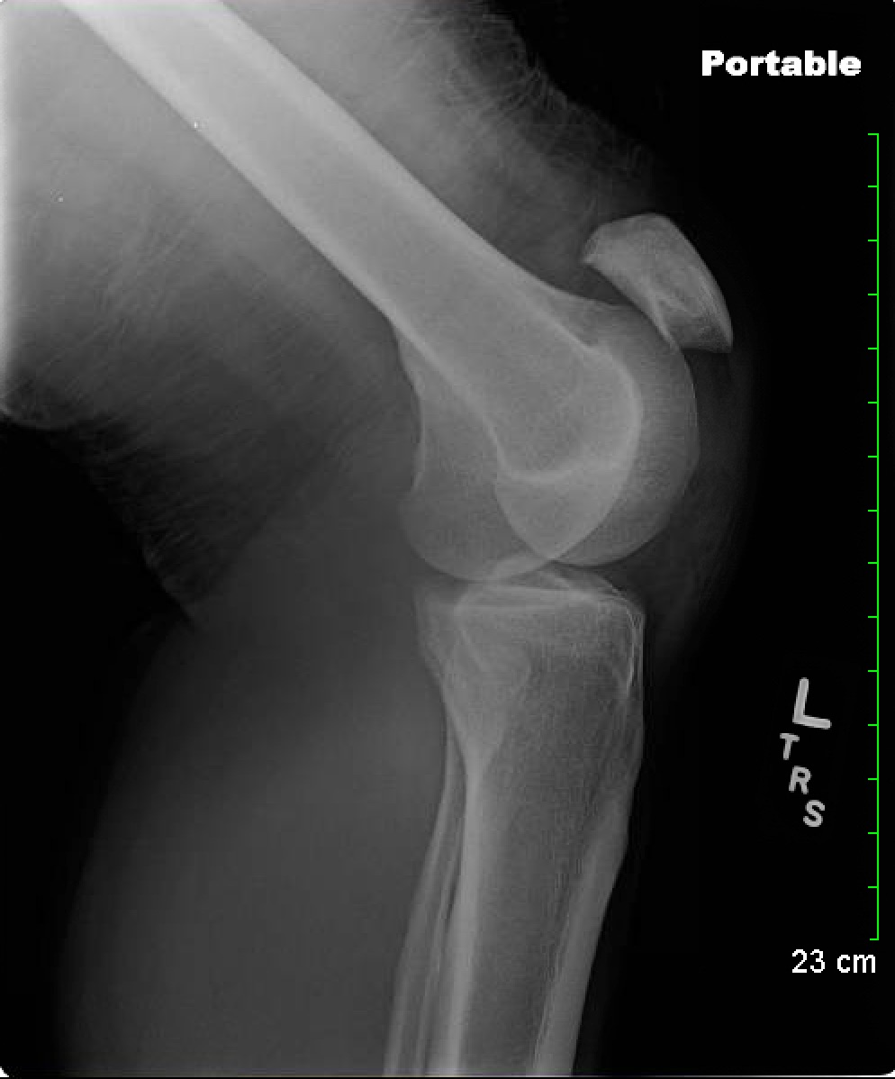
Figure 1: Xray of the left knee xray showing a high-riding patella.
The patient is a male in his 30s with no significant past medical history who presents with left knee pain and inability to ambulate after an assault. The patient works as a bouncer and shortly prior to arrival in the ED he fell backwards while restraining a rowdy bar patron. The patron fell directly on his left knee while his left foot was planted and his knee was flexed. On exam, the inferior border of the left patella can be palpated 5 cm above the tibial tuberosity. With his knee held in extension, he cannot lift his heel off the bed. He has no other knee instability. X-rays were obtained...
+ What is the diagnosis based on this X-ray?
X-ray of his left knee reveals a high-riding patella, also termed ‘patella alta’, confirming a complete patellar tendon rupture
Discussion and Management of Patellar Tendon Rupture
Patellar tendon rupture, as its name implies, is disruption of the tendon that connects the inferior patella to the tibial tuberosity. Trauma is the most common mechanism of rupture, occurring most frequently when a flexed knee resists a powerful contraction of the quadriceps muscle. Patients with systemic inflammatory disease, diabetes, chronic kidney disease, or chronic patellar tendinitis are at increased risk of atraumatic patellar tendon rupture.[1]
Physical exam in patients with patellar tendon rupture reveals a high-riding patella, diffuse swelling, and inability to ambulate on the affected leg. Additionally, patients have an impaired extensor mechanism such that they are unable to lift their heel with an extended knee. Our patient demonstrated all of these cardinal examination findings. Significant force is needed to rupture the patellar tendon, so care should be taken to evaluate patients for other ligamentous injury, as ACL and meniscal tears have been known to occur simultaneously with patellar tendon rupture.[2]
X-ray is the imaging modality of choice to diagnose patellar tendon rupture. Patella alta will confirm the diagnosis, and calcification of the patellar tendon inferior to the patella may indicate chronic patellar tendinitis. However, if the diagnosis cannot be established with x-ray and physical exam, MRI is indicated.[3] Ultrasound has also shown promise as an accurate imaging modality to diagnose patellar tendinopathy.[4] A patellar tendon rupture will be apparent by increased tendon thickness in the sagittal plane due to tendon retraction, as well as a wavy appearance of the tendon when comparing it to the uninjured knee.
Treatment of patellar tendon rupture is early primary operative repair, as delay can cause tendon contracture, scar tissue formation, and quadriceps muscle atrophy.[5] Therefore, urgent orthopedics follow-up must be established for patients. To prevent tendon contracture, the affected knee should be placed in a knee immobilizer and the patient should be non-weightbearing until they can be seen by an orthopedic surgeon.
Our patient was placed in a knee immobilizer and was discharged with urgent orthopedic surgery follow-up. He underwent successful open repair 2 weeks later. He has since followed up with orthopedic surgery and is healing well. He is currently able to ambulate without assistance.
Authored by Isaac Shaw, MD Posted by Grace Lagasse, MD


