Ultrasound Case of the Month - Sneaky Stones
/The Case:
A 25 y.o. male with a history of diabetes presents with a chief complaint of flank pain. The pain started earlier in the day as an "ache" in his right flank which wrapped around to his hip, and it was accompanied by diaphoresis and nausea. He had some straining with urination but no hematuria or dysuria. His pain does not seem to have any alleviating factors. He does endorse a strong family history of kidney stones with some family members having had lithotripsy and ureteral stent placement. The patient otherwise denies fever, chills, vomiting, chest pain, shortness of breath, hematuria, and diarrhea. His symptoms are treated with ketorolac and ondansetron while bloodwork and a urinalysis are pending. In the meantime, you decide to perform a bedside ultrasound.
What do you expect on ultrasound?
While on occasion you can directly visualize a very proximal or distal kidney stone, there are numerous other sonographic findings which may suggest nephrolithiasis, each with different sensitivities and specificities. These include hydronephrosis, acoustic shadowing, the twinkle sign, and absence of ureteral jets.
Proximal Renal Stones and Acoustic Shadowing
Proximal renal stones appear as bright echogenic foci, frequently with a posterior acoustic shadowing. Point of care ultrasound (POCUS) may be used not only to determine the presence of proximal stones, but also to determine the size and frequency of stones. May et al. evaluated stone size using ultrasound and acoustic shadowing and found that ultrasound was able to accurately measure the size of the stone within 2mm of the CT size estimation 73% of the time. Additionally, they found that larger stones were more likely to have a posterior acoustic shadow. Stones 5mm or larger were found to have acoustic shadow 89% of the time, while stones smaller than 5mm had an acoustic shadow only 53% of the time.
One of the more interesting types of renal stones one will encounter are staghorn stones. Given their proximal location, staghorn calculi are frequently visible when present. These stones, named for their staghorn like appearance, can be quite large and cause distortion of the renal pelvis. Additionally, the large posterior shadowing can obstruct ultrasonographic appearance of large portions of the kidney. These stones are commonly associated with urinary tract infections and need to be intervened on due to their ability to lead to urosepsis and renal failure. As we see below, point of care ultrasound will show what appears to be large confluent calculi extending into the renal calyces.

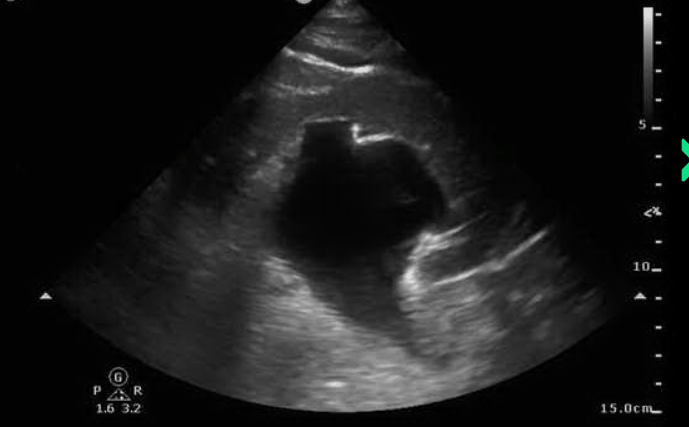
Proximal Kidney Stone

A Still of the adjacent Video demonstrating Stone at the junction of the Ureter and Kidney with Posterior Acoustic Shadowing
Hydronephrosis
Hydronephrosis refers to dilation of the renal pelvis on imaging and is graded from mild to severe. Sonographic hydronephrosis may be noted when a calculus results in distal obstruction to urine flow through the ureter. While a renal stone is one of the more common obstructive causes of hydronephrosis, they are not the only cause. There are also non-obstructive causes, such as vesicoureteral reflux, urinary tract infections, malignancy, pregnancy, or blood clot, which can lead to hydronephrosis. Renal calculi in a ureter will often result in unilateral hydronephrosis, whereas a more distal obstruction of any cause will result in bilateral hydronephrosis. As with any ultrasound, always compare to the contralateral side (when a contralateral is available) and remember to take the finding of hydronephrosis in clinical context.
Hydronephrosis is the most commonly used metric to assess for a clinically significant calculus. Wong et. al analyzed the prognostic value of hydronephrosis in a meta-analysis which included five studies. Using CT scan as a confirmatory study, their pooled findings demonstrated a sensitivity of 70.2% and a specificity of 75.4%. One study included in this meta-analysis evaluated 77 patients with renal colic, 28 out of which were found to have no hydronephrosis. Of those 28 patients without hydronephrosis, 0 required admission for a urological issue in the following 30 days. Two studies included in this meta-analysis demonstrated a specificity of 94.4% when either moderate or severe hydronephrosis was present. Additionally, four of the five studies showed that the sensitivity of hydronephrosis for nephrolithiasis was higher when the stones were at least 5mm in size, and another included study showed that the sensitivity of bedside renal ultrasound increased to 100% when patients had three or more stones, regardless of size.
While hydronephrosis is an incredibly helpful finding in the evaluation of renal colic, there are still documented cases of false-negative studies, especially in patients who have a partial obstruction or early obstruction. In acute cases of nephrolithiasis, the anatomical changes may not yet be evident when the patient arrives at the emergency department.

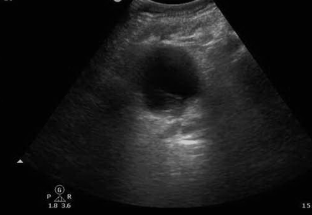
Moderate Hydronephrosis
Severe Hydronephrosis
Twinkle Sign

Twinkle Sign
More accurately described as an “artifact,” this interesting phenomenon appears as a focus of alternating colors (red and blue) behind an irregular reflective surface (like a kidney stone) on color Doppler, with the resultant image mimicking turbulent flow. Different theories have been evaluated over the past as to how to best acquire this artifact. Some posit placing the focal zone below the rough reflecting surface to allow better visualization of the twinkling compared to when the focal point is above the reflective surface (Ozan). Another technique is to reduce the number of ultrasound pulses emitted from the transducer, known as the pulse repetition frequency. The presence of a renal twinkle artifact has been shown to have a positive predictive value of 78% for the presence of a kidney stone being present anywhere in the kidneys on CT (Dillman). However, the same study showed that the sensitivity of a twinkle sign for a specific renal calculi was only 55%, with a false positive rate of 51%.
Ureteral Jets
Visualization of ureteral jets can be used as a surrogate for ureteral flow. The speed of the jet entering the bladder is rapid enough to be detected on Doppler. In patients who are well hydrated, this sudden burst of color can last for a few seconds and usually will occur twice or more per minute. If there is some obstruction of the ureter, it is expected that the duration of the jet would be reduced or the jet would be absent entirely. Unfortunately, there is very limited data evaluating the predictive value of this finding on ultrasound, and currently ureteral jets don’t have much prognostic value.
One prospective study evaluated ultrasound findings in a cohort of 77 people with renal colic, 13 of which required admission for urologic intervention, infection, or intractable pain. While hydronephrosis was found in all 13 of the patients who required admission, the absence of an ipsilateral ureteral jet was not found to be predictive of subsequent admission (Fields).
Another study, performed in Turkey, evaluated the peak velocity of a ureteral jet in predicting the likelihood of spontaneous stone passage. Seventy-four patients with an average stone size of 5.6mm were enrolled, and it was found that patients with a high ureteral jet peak velocity (> 15.25cm/s) had a high rate of stone passage (74.3%) (Ongung).
Ureteral Jet without Color Doppler

Ureteral Jet
Conclusion
Like everything in medicine, when it comes to patient workup and management, it is imperative to interpret findings in the clinical context of the patients’ illness. In the case of nephrolithiasis, POCUS has many findings which can help providers determine the presence of a clinically significant stone. Each of the findings listed above is a piece of the puzzle and it is important to recognize how they all fit together in getting to the final diagnosis.
Post by Dr. Eddie Irankunda
Dr. Irankunda is a PGY-3 in Emergency Medicine at the University of Cincinnati
Editing by Dr. Frederick and Dr. Baez
Dr. Frederick is a PGY-3 in Emergency Medicine at the University of Cincinnati.
Dr. Baez is an ultrasound trained Attending in Emergency Medicine at the University of Cincinnati.
References
Wong C, Teitge B, Ross M, Young P, Robertson HL, Lang E. The Accuracy and Prognostic Value of Point-of-care Ultrasound for Nephrolithiasis in the Emergency Department: A Systematic Review and Meta-analysis. Acad Emerg Med. 2018 Jun;25(6):684-698. doi: 10.1111/acem.13388. Epub 2018 Mar 25. PMID: 29427476.
Ozan E, Atac GK, Gundogdu S. Twinkling artifact on color Doppler ultrasound: an advantage or a pitfall? J Med Ultrason (2001). 2016 Jul;43(3):361-71. doi: 10.1007/s10396-016-0715-z. Epub 2016 Apr 28. PMID: 27126510.
Dillman JR, Kappil M, Weadock WJ, Rubin JM, Platt JF, DiPietro MA, Bude RO. Sonographic twinkling artifact for renal calculus detection: correlation with CT. Radiology. 2011 Jun;259(3):911-6. doi: 10.1148/radiol.11102128. Epub 2011 Apr 1. PMID: 21460031.
May PC, Haider Y, Dunmire B, Cunitz BW, Thiel J, Liu Z, Bruce M, Bailey MR, Sorensen MD, Harper JD. Stone-Mode Ultrasound for Determining Renal Stone Size. J Endourol. 2016 Sep;30(9):958-62. doi: 10.1089/end.2016.0341. PMID: 27393000; PMCID: PMC5031098.
Healy KA, Ogan K. Pathophysiology and management of infectious staghorn calculi. Urol Clin North Am. 2007 Aug;34(3):363-74. doi: 10.1016/j.ucl.2007.05.006. PMID: 17678986.
Klein I, Gutiérrez-Aceves J. Preoperative imaging in staghorn calculi, planning and decision making in management of staghorn calculi. Asian J Urol. 2020 Apr;7(2):87-93. doi: 10.1016/j.ajur.2019.07.002. Epub 2019 Jul 6. PMID: 32257800; PMCID: PMC7096669.
Fields JM, Fischer JI, Anderson KL, Mangili A, Panebianco NL, Dean AJ. The ability of renal ultrasound and ureteral jet evaluation to predict 30-day outcomes in patients with suspected nephrolithiasis. Am J Emerg Med. 2015 Oct;33(10):1402-6. doi: 10.1016/j.ajem.2015.07.014. Epub 2015 Jul 17. PMID: 26279392.
Ongun S, Teken A, Yılmaz O, Süleyman S. Can Ureteral Jet Flow Measurement Predict Spontaneous Passage of Distal Ureteral Stones? Urol Int. 2018;101(2):156-160. doi: 10.1159/000490498. Epub 2018 Jun 27. PMID: 29949810.


