Minor Care Series: Fingertip Injuries
/Fingertip Injuries:
Fingers and hands are essential to people for day to day living and work. It can be very distressing to sustain an injury to fingers. It is also a highly innervated area that can be uncomfortable and painful.
Many injuries can be managed in the emergency department, but it is also important to recognize when a hand surgeon should be involved. Maintaining function and aesthetics are both important in the treatment of fingertip injuries. We will go through a few cases and explore how we can treat fingertip injuries in the emergency department.
Classification:
Case 1:
16 year old male presents to the emergency department with chief complaint of fingertip injury. He is left handed and injured his right hand’s 4th digit while using a pocket knife breaking down boxes at his new job. Vitals stable and on exam you note no bone exposed from the injury. Approximately one centimeter of the distal finger on the volar aspect was affected and there is no nail bed involvement. There is moderate venous oozing and bleeding at the site of injury. The patient has trouble holding pressure at the site because of pain.
Anesthesia:
Stop the bleed! The patient likely won’t hemorrhage to death from his finger tip oozing, however it can add to the distress of the patient. Direct pressure is the best method to stop bleeding, but it can be very uncomfortable in these injuries due to the highly innervated site. A digital block after you finish a neurovascular exam of the hand can provide relief. Another option is soaking the the tip of finger in 1% lidocaine with epinephrine. This can be with a soaked gauze or in a small container with the solution.
Management:
This is an uncomplicated injury without bone exposure or nail bed involvement. Conservative management and secondary intention healing can be used in this patient. After irrigation, a non-adherent dressing can be placed in the emergency department. Educate the patient that this will take multiple weeks to heal. Keeping the site clean and using bandaids can help the healing area covered. Antibiotics are not indicated without bony exposure unless there is concern for a mechanism that can cause contamination or if patient has significant co-morbidities. Update tetanus as needed. Depending on job and compliance, consider a splint to help minimize movement and facilitate healing.
Case 2:
34 year old woman presents with the chief complaint of finger injury at work. This occurred within the past hour and she says the tip of her right middle finger was caught in a conveyer belt. On exam, there is a dorsal angulation to the injury and approximately 0.5 cm of exposed distal phalanx tuft.
Management:
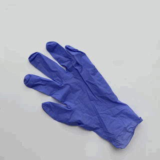
Creating a finger tourniquet from glove
An x-ray shows no fractures. Patient is up to date on tetanus. She is neurovascularly intact and the decision to rongeur the exposed bone and perform a primary closure. A digital block is performed for anesthesia of the finger. A finger tourniquet can be used to keep the site of injury non-bloody during this procedure. The finger of a medical examining glove can be cut to create a tourniquet as well. Please do not forget to remove the tourniquet if a critical patient in the emergency department arrives and requires a large amount of time away from your initial patient.
The rongeur can be used to trim the exposed bone before primary closure. This helps with wound healing and preventing complications from a hard piece of bone pushing against the closed soft tissue. Small pieces of bone should be trimmed and then the wound needs to be well irrigated to remove possible foreign bodies and contaminants. Primary closure can be performed with 5-0 or 6-0 nylon sutures. If necessary, a VY plasty can be performed to advance the flap over site of injury.
Dress the area with a non-adherent dressing and apply a bulky dressing to help protect the repaired finger. A splint can be considered for protection and healing as well. Though the evidence is sparse, experts generally recommend prophylactic antibiotics when bone is exposed. Follow up with a hand surgeon should be arranged and patient should watch for signs of infection. Though the evidence is sparse, experts generally recommend prophylactic antibiotics when bone is exposed. Follow up with a hand surgeon should be arranged and patient should watch for signs of infection.
Case 3:
52 year old male working with a table saw injured his right index finger. He reports the distal end was mangled in the saw. You are in a small community emergency department but do have most consulting services available by phone. He has a history of 2 pack per day smoking and also diabetes. On exam, you note a zone 3 injury and fractured bone at the site of injury. The injury is proximal to the flexor and extensor tendon insertions. You speak with the on-call hand surgeon and discuss with the patient about a revision amputation. Everyone agrees and patient can follow up in clinic after the procedure and being discharged home.
The distal interphalangeal joint will be disarticulated, preserving a short stump of the distal phalanx has little value. Apply traction, transect, and allow retraction of the profundus and extensor tendons. Rongeur the volar condyles of the head of middle phalanx to create a smooth contour. Identify and apply tension to the digital nerves so they can be transected away from the wound edge. This helps prevent painful neuromas. Close the wound with the palmer and dorsal skin, trim any excess skin to create ideal contour and appearance.
Post by James Li, MD
Peer Editing by Ryan LaFollette, MD
References:
Saraf, Sanjay, and Vk Tiwari. “Fingertip Injuries.” Indian Journal of Orthopaedics, vol. 41, no. 2, 2007, p. 163., doi:10.4103/0019-5413.32051.
Stearns, Dana A, and David A Peak. “Chapter 43: Hand.” Rosen's Emergency Medicine: Concepts and Clinical Practice, by Ron M. Walls et al., Elsevier, 2018.
Davenport, Moira. “Chapter 43: Arm and Hand Lacerations.” Tintinalli's Emergency Medicine: a Comprehensive Study Guide, 8th Edition, by J. Stephan. Stapczynski and Judith E. Tintinalli, McGraw-Hill Education, 2016.
Lemmon, Joshua A., et al. “Soft-Tissue Injuries of the Fingertip: Methods of Evaluation and Treatment. An Algorithmic Approach.” Plastic and Reconstructive Surgery, vol. 122, no. 3, 2008, p. 961., doi:10.1097/prs.0b013e318184d029.
“The Nuts & Bolts of Finger Amputation.” Emergency Physicians Monthly, epmonthly.com/article/the-nuts-a-bolts-of-finger-amputation/.
Jackson, Edward A. "The VY plasty in the treatment of fingertip amputations." American family physician 64.3 (2001): 455-458.
“Wheeless' Textbook of Orthopaedics.” Wheeless Online, www.wheelessonline.com/ortho/finger_tip_injuries.
“Fingertip Amputations & Finger Flaps.” Trauma - Orthobullets, www.orthobullets.com/hand/6060/fingertip-amputations-and-finger-flaps.
“Trick of the Trade: Fingertip Injuries.” ALiEM, 14 Jan. 2018, www.aliem.com/2011/06/trick-of-trade-fingertip-injuries/.
Guthrie, Kane. “Fingertip Injuries - Life in the Fast Lane Medical Education Blog.” Life in the Fast Lane • LITFL • Medical Blog, 25 Sept. 2017, lifeinthefastlane.com/insidious-injury-002/.
Fassler, Paul R. "Fingertip injuries: evaluation and treatment." JAAOS-Journal of the American Academy of Orthopaedic Surgeons 4.2 (1996): 84-92.


