Diagnostics and Therapeutics: The Who, What, Where, When and Why of Lumbar Punctures
/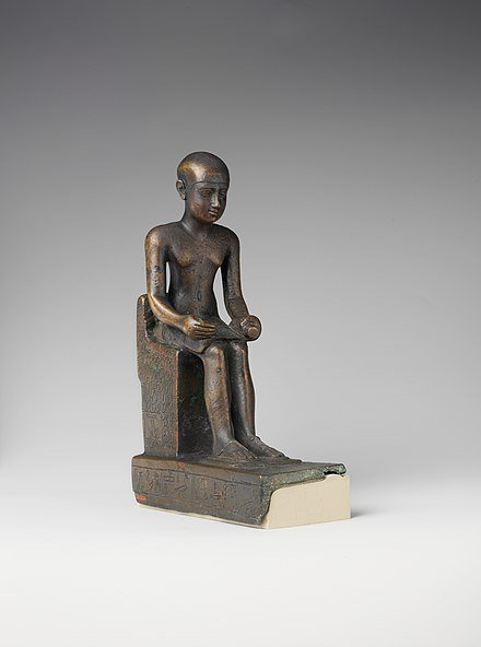
Imhotep was an egyptian chancellor and has been credited with the discovery of csf. Metropolitan Museum of Art, CC0, via Wikimedia Commons
The ancient Egyptian physician Imhotep is often credited with the discovery of cerebral spinal fluid (CSF) —over 5,000 years ago! (1) However, it wasn’t until the 1890s that purposeful, successful, and safe attempts to access this fluid were documented (2). Heinrich Quincke, of the eponymous Quincke needle, is often credited with this innovation and although his technique was perhaps not as sterile as that in use today, his second reported patient not only survived 3 lumbar punctures (LP), but symptomatically improved (1). The LP is now a standard procedure and in 2010 well over 135,000 LPs were performed in Emergency Departments throughout the US (3).
WHO - Who Needs an LP in the ED?
Patients with concern for infection (i.e., meningitis or encephalitis)
Patients with suspected or confirmed symptomatic Idiopathic Intracranial Hypertension (IIH)
Patients with concern for Subarachnoid Hemorrhage (SAH) in the setting of a negative head CT (after 6 hours of symptom onset)
Patients with other suspected inflammatory central nervous system pathology such as Guillain Barre Syndrome or Multiple Sclerosis (though the necessity of performing these studies in the ED may be up for debate)
WHAT - What Do You Do?
1. Position the patient (see “where” below).
2. Palpate for the posterior superior iliac crest, which approximates the level of the L4 spinous process.
3. Sterilize the needle insertion site (L3-L4 or L4-L5) and the surrounding area with chlorhexidine or betadine.
4. Don sterile gloves and sterilely drape the patient.
5. Use a 25-27 gauge needle to anesthetize the skin by creating a wheal with lidocaine. Next, anesthetize a tract into the intended LP site by inserting the needle midline, parallel to the stretcher, and angled slightly towards the umbilicus as this is the same course your spinal needle should follow.
6. Set up your collecting tubes and manometer.
7. Insert the spinal needle along the tract with the bevel parallel to long axis of spine until you enter the subarachnoid space. The layers you will pass through are the skin, the subcutaneous layer, muscle, the supraspinous ligament, the interspinous ligament, the ligamentum flavum, the epidural space, the dura, and then you are in! You may remove the stylet multiple times to check if CSF is flowing. Once fluid begins to trickle out, collect the fluid into 4 numbered tubes. For a typical LP in an adult with no contraindications, collect least 1cc, preferably 2-3cc’s, per tube.
8. If measuring opening pressure, this must be done prior to fluid collection, and must be performed with the patient in lateral decubitus position. The patient’s legs must be straightened for accurate result. This video demonstrates how to set up the manometer.
9. Once CSF collection is complete, re-insert the stylet, remove the needle, and apply a bandage to the entry site.
Troubleshooting – ideas to ponder if CSF remains elusive
Is your needle midline? If not, reposition (you will likely need to withdraw the needle somewhat to correct your path).
Is your needle still parallel to the stretcher? If not, correct your trajectory (you will likely need to withdraw the needle somewhat to correct your path).
Are you hitting bone? If you are hitting bone at a shallow depth (1-2cm), you may be encountering the spinous process and may benefit from inserting 1 cm superior or inferior to your previous attempt. If you are hitting bone at a deeper depth (2-4cm), your trajectory may be skewed laterally and you may be encountering the lamina. Attempt to redirect your needle medially. This helpful video demonstrates these concepts and demonstrates how to correct further attempts.
WHEN - When NOT to do an LP
Contraindications - Absolute
Infection (i.e. cellulitis) overlying the needle insertion site
Spinal epidural abscess
Contraindications - Relative
Thrombocytopenia (< 20,000 - 50,000), coagulopathy, INR > 1.6, or on oral anticoagulation
Known trauma to the insertion site
Known intracranial process causing mass effect, CT showing midline shift/brainstem herniation, compression/obliteration of suprachiasmatic and basilar cisterns/4th ventricle, signs of increased ICP
If LP positioning would cause respiratory compromise
WHEN - When Should You Get a CT first?
(And if concerned for bacterial meningitis, give antibiotics before the CT)
There are a variety of guidelines regarding which patients require a CT prior to an LP. As may be seen in the table below, there are several themes as well a range of stringency.
| Swedish (5) | UK (6) | ESCMID (7) | IDSA (8) |
|---|---|---|---|
| Signs of cerebral mass lesions (Focal neurologic deficit, new onset epileptic seizures if not associated with clinical picture indicating acute bacterial meningitis, >4 days duration of cerebral symptoms) | GCS ≤ 12 | GCS<10 | Abnormal level of consciousness |
| Ongoing or impending cerebral herniation (Unconsciousness plus 1 or more of the following: rigid dilated pupils, increasing blood pressure and bradycardia, disturbed breathing patter, opisthotonus, loss of all reactions) | Focal neurologic deficit | Focal neurologic deficit | Focal neurologic deficit |
| Ongoing epileptic seizures | Papilledema (though inability to view fundus not considered contraindication to LP especially in those with short symptom duration) | Severely immunocompromised state | Immunocompromise |
| Papilledema (ophthalmoscopy is not mandatory before LP with acute onset and short duration of symptoms) | Continuous or uncontrolled seizures | New onset seizures | Papilledema |
| History of CNS disease (mass, stroke, focal infection) | |||
| New onset seizure (within 1 week of presentation) |
WHERE - Landmark and Ultrasound Guided
Positioning
Patients may sit or lie in the lateral decubitus position. Find the posterior superior iliac crest and draw an imaginary line across the back (Tuffier’s line) – where this line crosses midline is the L4 spinous process. Opening pressure can only be measured in the left lateral decubitus position with the legs straightened (at least with the typical manometer). Any positioning that widens the gap between the spinous processes (such as arching the back, curling the back, lifting the legs if sitting), may ease the path to the subarachnoid space.
Tips for the seated position:
Try having the patient place their feet on a footstool to elevate their legs
Have the patient curl forward as much as possible to ensure the back is rounded (someone to help support the patient may be quite helpful, and if not, the patient can rest their arms on their knees or use a tray for support)
Tips for the lateral decubitus position:
Ensure shoulders are even and the back is perpendicular to the stretcher
Bend the patient’s knees upwards/towards the abdomen/chest (if measuring opening pressure the legs can be straightened after entering the space

Lumbar puncture positioning both lying and sitting - courtesy of https://almostadoctor.co.uk/encyclopedia/lumbar-puncture

Baby LP positioning while sitting - courtesy of https://garystockbridge617.getarchive.net/amp/media/spinal-tap-being-performed-on-a-newborn-baby-to-detect-presence-of-infection-cd2a85
Ultrasound-assisted approach
Ultrasound may be useful in patients for whom a solely landmark based approach may be more difficult, including for example, obese patients, patients with ankylosing spondylitis, kyphoscoliosis, previous lumbar surgery, degenerative disk disease, or osteoarthritis.
1) Find the anatomic midline with the probe in the transverse orientation and identify the spinous processes, center this, and mark this on the patient. The curvilinear probe may be most helpful here given its wide footprint, but the linear probe can also be utilized in patients with a lower BMI.
Transverse view of lumbar spinous process
The spinous processes are superficial structures visible in the midline. If you slide slightly laterally, you will see the lamina that are further in the far field of the screen.
2) Switch to the longitudinal orientation and identify the spinous processes with the interspinous space in between, center this, and mark the patient at this space to identify the midline. Be sure to distinguish the superficial spinous processes from the lamina (which are slightly lateral and will be further into the far field).


The sacrum is visible on the right side of the screen and provides an means to determine spinal levels if landmarks are not easily palpable
3) Insert the needle where these lines cross. It is most helpful to do the ultrasound immediately before needle insertion, as movement of the patient may shift cutaneous landmarks from underlying bony structures.
If needed, you can use US to identify L3/4/5 (use the probe in the longitudinal axis) by finding the sacrum. You can also estimate the depth to the subarachnoid space by identifying the ligamentum flavum and measuring the distance from this structure to the skin surface.
This resource offers additional information on ultrasound assisted LP’s.
WHY - Why Not?
Potential complications include post LP headache, nerve injuries, hematomas (spinal, retroperitoneal), subarachnoid epidermal cysts, infection, and herniation. Most of these are quite rare when following proper protocol, with the exception of the post-LP headache.
POST BY Chloe Knudsen-Robbins, MD
Dr. Knudsen-Robbins is a PGY-1 in Emergency Medicine at the University of Cincinnati.
EDITING BY Maksim Kletsel, MD AND Arthur Broadstock, MD
Dr. Kletsel is a PGY-4 and Chief Resident in Emergency Medicine at the University of Cincinnati.
Dr. Broadstock is a Clinical Instructor and Ultrasound Fellow in Emergency Medicine at the University of Cincinnati and an Assistant Editor of TamingtheSRU.
REFERENCES
1. Zambito Marsala, S., Gioulis, M., & Pistacchi, M. (2015). Cerebrospinal fluid and lumbar puncture: the story of a necessary procedure in the history of medicine. Neurological sciences : official journal of the Italian Neurological Society and of the Italian Society of Clinical Neurophysiology, 36(6), 1011–1015. https://doi-org.uc.idm.oclc.org/10.1007/s10072-015-2104-6
2. Frederiks, J. A., & Koehler, P. J. (1997). The first lumbar puncture. Journal of the history of the neurosciences, 6(2), 147–153. https://doi-org.uc.idm.oclc.org/10.1080/09647049709525699
3. Vickers A, Donnelly JP, Moore JX, Barnum SR, Schein TN, et al. (2018) Epidemiology of lumbar punctures in hospitalized patients in the United States. PLOS ONE 13(12): e0208622. https://doi.org/10.1371/journal.pone.0208622
4. Glimåker M, Sjölin J, Åkesson S, Naucler P. Lumbar Puncture Performed Promptly or After Neuroimaging in Acute Bacterial Meningitis in Adults: A Prospective National Cohort Study Evaluating Different Guidelines. Clin Infect Dis. 2018;66(3):321-328. doi:10.1093/cid/cix806
5. Glimåker, M., Johansson, B., Bell, M., Ericsson, M., Bläckberg, J., Brink, M., Lindquist, L., & Sjölin, J. (2013). Early lumbar puncture in adult bacterial meningitis--rationale for revised guidelines. Scandinavian journal of infectious diseases, 45(9), 657–663. https://doi.org/10.3109/00365548.2013.799289
6. McGill, F., Heyderman, R. S., Michael, B. D., Defres, S., Beeching, N. J., Borrow, R., Glennie, L., Gaillemin, O., Wyncoll, D., Kaczmarski, E., Nadel, S., Thwaites, G., Cohen, J., Davies, N. W., Miller, A., Rhodes, A., Read, R. C., & Solomon, T. (2016). The UK joint specialist societies guideline on the diagnosis and management of acute meningitis and meningococcal sepsis in immunocompetent adults. The Journal of infection, 72(4), 405–438. https://doi.org/10.1016/j.jinf.2016.01.007
7. van de Beek, D., Cabellos, C., Dzupova, O., Esposito, S., Klein, M., Kloek, A. T., Leib, S. L., Mourvillier, B., Ostergaard, C., Pagliano, P., Pfister, H. W., Read, R. C., Sipahi, O. R., Brouwer, M. C., & ESCMID Study Group for Infections of the Brain (ESGIB) (2016). ESCMID guideline: diagnosis and treatment of acute bacterial meningitis. Clinical microbiology and infection : the official publication of the European Society of Clinical Microbiology and Infectious Diseases, 22 Suppl 3, S37–S62. https://doi.org/10.1016/j.cmi.2016.01.007
8. Tunkel, A. R., Hartman, B. J., Kaplan, S. L., Kaufman, B. A., Roos, K. L., Scheld, W. M., & Whitley, R. J. (2004). Practice guidelines for the management of bacterial meningitis. Clinical infectious diseases : an official publication of the Infectious Diseases Society of America, 39(9), 1267–1284. https://doi.org/10.1086/425368


