
Riding the Waves: End-Tidal CO2 Monitoring
End-Tidal CO2 monitoring has a variety of uses in the Emergency Department. Whether used diagnostically or for monitoring of a patient’s physiology, clinicians must possess an understanding of the information that you can gather from EtCO2 waveform tracings. Knowing how to interpret the waveforms makes EtCO2 much more than a number, allowing the clinician to gain insight into minute to minute changes in a patients physiological state.

End-Tidal CO2 monitoring has a variety of uses in the Emergency Department. Whether used diagnostically or for monitoring of a patient’s physiology, clinicians must possess an understanding of the information that you can gather from EtCO2 waveform tracings. Knowing how to interpret the waveforms makes EtCO2 much more than a number, allowing the clinician to gain insight into minute to minute changes in a patients physiological state.
Respiratory Physiology
Fortunately, to understand waveform capnography, we don’t need an in-depth knowledge of respiratory physiology. The most important things to remember are: 1) ambient air contains effectively 0% carbon dioxide whereas exhaled air contains about 4.4% carbon dioxide, and 2) the dead space in your airway does not participate in gas exchange.
Knowing this, we can take a look at an end-tidal waveform and understand discrete parts of the wave and what they represent throughout the respiratory cycle.

Phase 0: This part of the curve in inspiration. During this phase, the CO2 value should be 0 as the capnometer is detecting only ambient air which should contain effectively 0 CO2.
Phase 1: This part of the curve is the earliest part of expiration. This initial expiration contains only dead space gas that has not mixed with any gas from the conducting airway, so there is no increase in CO2. This plateau should have a gentle upslope or remain flat.
Phase 2: This transitional part of the curve still represents early expiration. During this phase gas from the upper airways that is low in CO2 mixes with gas from the conducting airways. The amount of CO2 increases in the expired air increases as you approach the alveolar plateau.
Phase 3: This part of the curve is called the alveolar plateau. It represents the steady-state diffusion of CO2 in the alveoli. The peak of this plateau is the end-tidal CO2. The angle of change between Phase 2 and Phase 3 of respiration is referred to as the alpha angle.
Phase 0: As expiration ends, the capnometer again senses ambient air with almost 0 CO2, and the waveform instantaneously drops to 0.

Knowing this, the waveform can give us a lot of useful information. In addition to the numerical end-tidal CO2 which is a function of metabolic rate and cardiac output, we get useful information about air movement in the airways and the length of time of inspiration and expiration. This means that the waveforms can offer assistance with the diagnosis of lung pathology.

The shape of this waveform represents an asthma exacerbation or bronchospasm. As bronchial constriction causes worsening obstruction the slope of Phase 2 gets smaller as it takes a longer time to clear out dead space gas. Further, there is no true alveolar plateau as the dead space has not completely cleared by the end of expiration. The alpha angle disappears, giving a classic sawtooth appearance.

This waveform represents emphysema. Recall that in emphysema the pathophysiology is related to the destruction of alveoli and increased compliance of the lungs. Because of the alveolar destruction, the surface for gas exchange is decreased, and the alveolar plateau becomes downsloping.
While these features may not be necessary to manage patients, it allows us to explore and understand the pathophysiology and how it aligns with what we see on our monitors. However, there are times when the use of end-tidal CO2 monitoring is critical to patient management.
Intubation
End-tidal monitoring is the most accurate method for immediate confirmation of endotracheal tube placement. Breath sounds, fogging or condensation in the ET tube, and chest rise may all be present in esophageal intubations. Whereas the capnogram for esophageal intubation will quickly drop to a flatline.
Capnography is also particularly useful in the transport of intubated patients. Transport is associated with a higher risk of tube displacement. Continuous monitoring allows for rapid recognition of tube displacement.
Cardiac Arrest
As mentioned before, tidal CO2 is a function of metabolic rate and cardiac output, so capnography can be utilized during CPR to monitor the effectiveness of chest compressions. Positive waveforms should be seen with high-quality compressions, and a gradual decline in end-tidal CO2 should be a signal that a fresh provider should perform compressions. An abrupt increase in end-tidal is an indicator of the return of spontaneous circulation (however this can also represent recent administration of bicarbonate).
Procedural Sedation
There are a number of studies that prove that capnography during sedation detects apnea earlier, leads to earlier intervention, and decreases morbidity and mortality when compared to pulse oximetry monitoring.
For some additional background, take a look and listen at our previous posts/podcasts on EtCO2.
References
Long, Brit, Alex Koyfman, and Michael A. Vivirito. "Capnography in the emergency department: a review of uses, waveforms, and limitations." The Journal of emergency medicine 53.6 (2017): 829-842.
Whitaker, D. K. "Time for capnography–everywhere." Anaesthesia 66.7 (2011): 544-549.
Kelly, John J., et al. "Use of tube condensation as an indicator of endotracheal tube placement." Annals of emergency medicine 31.5 (1998): 575-578.
Tobias, Joseph D., Amy Lynch, and Jeremy Garrett. "Alterations of end-tidal carbon dioxide during the intrahospital transport of children." Pediatric emergency care 12.4 (1996): 249-251.
Neumar, Robert W., et al. "Part 1: executive summary: 2015 American Heart Association guidelines update for cardiopulmonary resuscitation and emergency cardiovascular care." Circulation 132.18_suppl_2 (2015): S315-S367.
Waugh, Jonathan B., Chad A. Epps, and Yulia A. Khodneva. "Capnography enhances surveillance of respiratory events during procedural sedation: a meta-analysis." Journal of clinical anesthesia 23.3 (2011): 189-196.
Godwin, Steven A., et al. "Clinical policy: procedural sedation and analgesia in the emergency department." Annals of emergency medicine 63.2 (2014): 247-258.
Lightdale, Jenifer R., et al. "Microstream capnography improves patient monitoring during moderate sedation: a randomized, controlled trial." Pediatrics 117.6 (2006): e1170-e1178.
Authorship
Written by Dave Wilson, MD, PGY-1, University of Cincinnati Department of Emergency Medicine
Peer Review and Editing - Jeffery Hill, MD MEd, Associate Professor, University of Cincinnati Department of Emergency Medicine
Cite As
Wilson, D. Hill, J. Riding the Waves: End-Tidal CO2 Monitoring. TamingtheSRU. www.tamingthesru.com/blog/core-content/riding-the-waves-end-tidal-co2-monitoring. 7/2/2023.

Grand Rounds Recap 6.21.23
This week’s grand rounds starts off strong with the last Morbidity and Mortality of the year presented by Dr. Zalesky. This included multiple stimulating cases including meningitis, procedural safety, acute aortic syndromes, hemophilia, electrical storm, and euglycemic DKA. Finally, we wrap up the day with a lecture on the science behind wellness with Dr. Martella.
Morbidity and mortality WITH dr. zalesky
Meningitis
Meningitis is an extremely difficult diagnosis with almost no clinical findings that can aid in the diagnosis outside of suspicion and a LP
Seizures can be a presenting sign of infections, often without fevers or leukocytosis
Of patients with seizures who underwent LP, 22% had an infection (with about half of these patients without any other clinical signs of infection)
Hydrocephalus is a rare complication of bacterial meningitis, which often portends a poor outcome
There is scant evidence for platelet thresholds for LP, but the agreed-upon level is 50k
Procedural Safety
Retained guidewires are considered a never event
Based on a study on guidewire retention, there is no obvious identifiable risk factor
Checklists and a “wire count” are best practice
Acute Aortic Syndromes
Acute aortic syndromes are made up of dissections, aneurysms, penetrating atherosclerotic ulcers, and intramural hematomas
PAU, when symptomatic, can carry a mortality of up to 30%
Intramural hematomas will progress to aortic dissections 16-40% of the time
AAA >8cm have up to a 50% rupture rate at 1 year
Any concern for aortic pathology associated with new pain should be treated with a high level of concern
Hemophilia
Factor Replacement should be given at the suspicion of bleeding before any diagnostics are completed
When in doubt, administer factor immediately
Replacement should be administered before transport when at all possible
Hemlibra acts prophylactically and dramatically reduces bleeding events for patients with Hemophilia A
Give full-factor replacement for any critical bleeds
Factor VIII: full replacement dose 50 units/kg
Factor IX: full replacement dose 100 units/kg
If you do not have factor, can try cryo, DDAVP, FFP, aPCC, and aVII
Electrical Storm
Three episodes or more of sustained ventricular arrhythmia over the course of 24 hours
Management
Early aggressive antiarrhythmics
Amiodarone: first line
Procainamide: best at stopping VT, however lots of contraindications
Lidocaine: best agent for monomorphic scar-related VT
Magnesium: first line for torsades
Early beta blockade is important
Initial esmolol bolus followed by gtt
Early and aggressive anxiolysis & sedation
Dexmedetomidine recommended
Patient will likely ultimately require intubation and sedation with propofol
Don’t neglect the ICD and EP assistance as you may be able to utilize overdrive pacing
Hemodynamic support is a bridge to treatment
Euglycemic DKA
SGLT2 Inhibitors “–glifozin”: block the resorption of glucose leading to glucose loss in the urine while also increasing ketosis
SGLT2i are becoming more commonly used in T1DM, T2DM, HFpEF, and HFrEF
Patients are prone to ketosis and ketoacidosis, especially if a trigger is present
Patients on SGLT2i require an extra moment of consideration for euglycemic DKA
Respect significant derangements in bicarb levels or VBG abnormalities
Wellness: Behind the curtain WITH dr. Martella
Anxiety, depression, and suicide rates in resident physicians is high; this ultimately lead to ACGME mandating that wellness is a core requirement of residency programs
Wellness is the active pursuit of activities, choices, and lifestyles that lead to a state of holistic health
Domains of wellness:
Physical
Mental
Spiritual
Emotional
Social
Environmental
Many residencies have been implementing strategies to maintain residency wellness. While these programs differ, many have shown improvement in perceived wellness among residents.
Grand Rounds Recap 6.7.23
This week Dr. Diaz starts off with a challenging case of massive upper GI bleed managed with balloon tamponade. We then moved into a case follow-up with profound electrical storm and recurrent ventricular arrhythmias secondary to a STEMI. Following this, we took a deep dive into waveform capnography regarding normal physiology and alterations with lung pathology with Dr. Wilson. Next, we had an exciting CPC showdown where Dr. Bryant successfully diagnosed Dr. Haffner’s case of valproic acid toxicity presenting with hyperammonemic encephalopathy. We closed with Dr. Wosiski-Kuhn outlining the difficulty of intubation in a patient with DKA and severe metabolic acidosis.
Taming the sru WITH dr. diaz
Case: Middle aged patient with esophageal varices, recently banded presents for large volume hematemesis in hypovolemic shock. She required MAC line placement, MTP activation and transfusion. She was intubated for airway protection and the decision was made to perform balloon tamponade with placement of a Minnesota Tube.
Balloon Tamponade for Massive UGI Bleed:
- Minnesota tube vs Blakemore tube: MT has extra isolated esophageal aspiration port, as well as more air in gastric balloon
- Indications: acute UGI (mostly variceal) unresponsive to medical therapy
- Contraindications: known esophageal stricture, recent surgery at the GE junction
- Complications: esophageal perforation/erosions, aspiration
Steps:
Intubate the patient. Recommend using rocuronium to give you time with the patient paralyzed.
Insert 3-way stopcocks to inflation ports
Test balloon in water to evaluate for leaks and deflate entirely
Prep tube: consider placing in bucket of ice to stiffen for easy placement, lubricate with jelly and consider bougie insertion into the distal port to ease guidance
Insert under direct visualization with video laryngoscope to 50cm
Inflate 50cc of air into gastric balloon
Obtain an xray to ensure gastric balloon is below diaphragm
Fully inflate to 500cc and clamp balloon port
Pull back until some resistance is felt, usually ~40cm, secure with ETT holder or tie Minnesota tube with 1-2lbs of tension to ensure appropriate placement
Aspirate from esophageal port and potential need for intervention
If planning to intervene, use cufflator to inflate balloon to 30mmHg
See AirCare Taming the SRU article: Balloon Tamponade of Variceal Hemorrhage
R4 Case Follow-up WITH dr. gressick
Case: Patient presented post-arrest from recurrent VT/VF secondary to STEMI in electrical storm.
Electrical storm: 3+ sustained episodes of VT, VF or appropriate ICD shocks during a 24 hour period
- Causes: drug toxicity, electrolyte disturbance, new/worsened heart failure, acute myocardial ischemia, thyrotoxicosis, QT prolongation
- Treatment: high-quality CPR if under arrest as well as appropriate use of cardioversion/defibrillation, amiodarone, magnesium, lidocaine
- Attenuation of sympathetic drive: esmolol (or other BB), avoid epinephrine, u/s-guided stellate ganglion blockade
- Treat the underlying cause
r1 clinical diagnostics: waveform capnography WITH dr. wilson
End-tidal CO2: Information we are getting: numerical CO2, which has to do with our metabolic rate and cardiac output
- Time of inspiration/expiration
- Information about air movement

Phase 0: inspiration
- CO2 should be 0 due to low amount of CO2 in normal air
Phase 1: early expiration, purely deadpace
- The air inside the patient rushes out of them driven by the spring-like recoil of the chest wall and lung parenchyma
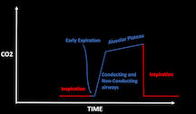
Phase 2: mixing dead space and conducting airways
- Gas from the upper airways (poor in CO2) slowly gives way to mixed gas from the lower airways (rich in CO2)
- Alpha angle: transition point between airway gas and alveolar gas; once dead space has emptied, the remaining gas exchange is a passive mixing of the gas in the tubing and gas inside the alveoli
Phase 3: approach equilibrium, alveolar plateau
Phase 0: inspiration
Specific scenarios: Asthma exacerbation: sawtooth slope due to obstruction in bronchi; the dead space does not finish emptying by the time the next inspiration begins leading ot loss of the transition angle
Mechanical airway obstruction: inspiratory and expiratory flow will be affected; their is a less steep transition to inspiration demonstrating that the obstruction can’t be overcome even when a patient is ventilated
Emphysema: aleveolar slope is reversed; due to poor gas exchange and abnormally increased lung compliance, the alveolar gas exchanges very rapidly. The part of the curve that represents the arterial CO2 is the early peak, not the end-tidal value. After, gas in the ventilator tubing diffuses backwards into the patient, at which point an equilibrium between the higher CO2 in the patient and the lower CO2 in the ventilator circuit is reached which results in a gradual drop of the CO2 concentration
Pigtail capnogram: seen in poor lung compliance, as well as some pregnant women and obese patients. Sudden peak of pre-inspiratory expired CO2 due to sudden airway closure. The last few milliliters of CO2-rich gas is expired before the collapse of the lung parenchyma occludes the bronchi and puts an end to the escape of gas.
Clinical pathologic case WITH drs. haffner and bryant
Valproic Acid Toxicity with Hyperammonemic Encephalopathy
Valproic acid: antiepileptic agent
Blocks voltage-gated sodium channels
Increases brain GABA concentrations
No direct effect on GABA(A) receptors
Complex mechanism
Overdose can cause hyperammonemia
Small volume of distribution, at therapeutic levels, VPA is >80% protein bound
Serum levels:
50-100 ug/ml: therapeutic
>180: mental impairment
>450: serious intoxication
>850: coma likely
Diagnosis
Ammonia level, valproic acid level
Electrolytes and liver function tests
CK
Management
Supportive care: ABCs
Treat the source: stop valproic acid, consider charcoal, consider hemodialysis
Provide antidote: L-carnitine
Loading: 100mg/kg over 30 minutes
Maintenance: 15mg/kg every 3-4 hours
Consider naloxone, carbapenems (reduce effectiveness of valproic acid)
taming the sru WITH dr. wosiski-kuhn
Case: Critically ill patient presenting with profound metabolic acidosis from DKA who unfortunately necessitates intubation for airway protection and severe acute hypoxic respiratory failure.
Intubating DKA: try to avoid it if possible; high risk of cardiovascular collapse and arrest due to difficulty matching metabolic demand on ventilator and apneic period leads to CO2 retention and worsening overall acidosis
Correct hypovolemia; start with MAP >75
Attempt to correct metabolic derangements
Be prepared for emesis
Apneic ventilation with BVM
Large ETT to minimize airway resistance
As soon as ETT is secured, restart your hyperventilation to prevent arrest from CO2
Tidal volume 8cc/kg
High RR
Rocuronium- assists with ventilator synchrony
Consider giving bicarbonate prior to intubation if HCO3 <10

Mastering Minor Care: Dog Bites
Don’t worry I’m a DOG-ter! Join Dr. Yates for a deep dive into dogbites covering which wounds to repair, when to consider antibiotics and whether a rabies vaccine is indicated.
In the United States, approximately 4.5 million people are bitten by dogs annually [1]. Although not every person seeks health care for these injuries, the majority of those that do end up in the emergency department. In 2008, this led to greater than 316,000 emergency department (ED) visits and about 9,500 hospitalizations [1]. There are multiple factors to consider when managing dog and mammalian bites in the ED, including closure, prophylactic antibiotic administration, and rabies post-exposure prophylaxis. Let’s take a deeper dive into the evidence.
To close or not to close?
One of the major questions when faced with a patient who has any mammalian bite in the ED is “am I going to repair that?” While the evidence is extremely limited, there are some consistent themes to help guide your management. Wounds on the face and head - due to significant improvement in cosmesis - should be primarily closed [2,3]. Also in the literature, there is often discussion of whether or not primary closure affects risk of infection. The consensus from several studies including a meta-analysis revealed that there was no increased risk from primary closures [3,4,5], even if antibiotics were not given.
However, IDSA guidelines still only recommend primary closure on the face [2]. Additionally, there seems to be significantly increased risk of infection with wounds presenting to the ED more than eight hours after injury, so caution should be used if evaluating these wounds for repair [3]. With gaping wounds that are not on the face, and particularly on the hand with highest likelihood of infection, primary closure can be determined by a shared decision-making conversation with the patient.
Bottom line: Dog bite wounds should always be primarily closed on the face for cosmetic purposes, but caution and shared decision making should be used on all other wounds, particularly those presenting greater than 8 hours from injury, due to uncertain increased risk of infection.
I heard dog mouths are dirty. I should put them on antibiotics, right?
Table 1: antibiotic regimens
“Am I going to give antibiotics?” should be the next question answered in any visit for a dog bite. While Amoxicillin-Clavulanate is the first-line antibiotic for 3-5 days after injury, alternate regimens are available for broader coverage (Table 1) [2]. This is due to studies showing mixed aerobes and anaerobes in isolates of wounds from dog bites [6]. But before you sign that prescription, some ED physicians question if antibiotics should be prescribed at all. There is no strong consensus as to whether dog bite lacerations lead to an increased risk of infection compared to other wounds [7]. One meta-analysis reported a rate of infections after dog bites of 3-46% [8] versus 2-5% in simple laceration repairs in the ED [9,10]. However, there have been no studies statistically powered well enough regarding dog bite lacerations to confirm or dispute this claim.
Table 2. Considerations for antibiotics.
The meta-analysis most frequently cited showing benefit for prophylactic antibiotics in dog bite wounds was performed in 1993 [8]. Although there was large study heterogeneity, it found a statistically significant relative risk for infection of 0.56 with a number needed to treat of 14 patients. More recently, a Cochrane review which consisted almost entirely of the same studies as the original Cummings meta-analysis, found no significant difference in rate of infections with prophylactic antibiotic use [11]. Finally, a randomized-control trial found that prophylactic antibiotics for dog bite wounds did not statistically change infection outcomes and was not cost-effective, recommending that prophylactic antibiotics only be used in high-risk wounds [12]. One study characterized puncture wounds and those with primary closure as the wounds at most increased risk for infection [13], while additional studies have indicated wounds on the hand [9] and wounds seen >8 hours after injury [3] as having the highest risk of infection. Table 2 has been included to summarize these results, most consistent with the current IDSA guidelines [2].
Bottom line: Antibiotics have not been shown to be significantly beneficial in most studies, and consideration to a patient's co-morbidities may be more helpful in judicious use of antibiotics rather than prophylactically prescribing to all.
What about rabies?
The final consideration is “do I have to give them rabies prophylaxis?” In the United States, carnivorous wild animals and bats are the most common source of transmission to humans. In 2006, only 79 dogs were found to have rabies in the United States [14]. If a patient sustains a domestic dog bite, that dog (if able) should be observed for 10 days even if the bite was provoked and the animal is vaccinated. If that animal develops any illness consistent with rabies, the patient should receive rabies post-exposure prophylaxis. If there is high suspicion that the animal is rabid, such as an ill animal with an unprovoked bite, then post-exposure prophylaxis should be given immediately. If the animal cannot be observed, this can be discussed with the patient and the public health department on the risks and benefits of immediate versus delayed treatment [14].
Post-exposure rabies prophylaxis involves both the Human Rabies Immune Globulin (HRIG, HyperRab) and the rabies vaccine (Rabavert). HRIG should not be given if the patient has previously received the rabies vaccines. HRIG should be administered at a 20 IU/kg dose intramuscularly. If possible, this should be administered into and around the wound(s) with the remainder given distant to the vaccine [15]. The first 1.0mL of the rabies vaccine, either the human diploid cell vaccine or the purified chick embryo cell vaccine, should be administered in the deltoid (patients >/= 2 years old) on the first day of presentation and additionally given on days 3, 7, and 14 for a total of four doses [15]. A 5th dose on day 28 should be given for immunocompromised people.
In patients younger than 2, this should be given in the anterolateral thigh. The gluteus should never be used because observations suggest administration in this area results in lower neutralizing antibody titers. If HRIG was not administered when vaccination was begun, it can be administered up to 7 days after the administration of the first dose of vaccine. Beyond the 7th day, HRIG is not recommended since an antibody response to the vaccine is presumed to have occurred. Patients should ideally avoid corticosteroids, antimalarials, and other immunosuppressants during post-exposure therapy as this can inhibit the immune response. The vaccine is safe in pregnancy and breastfeeding.
Bottom line: Rabies post-exposure prophylaxis should be discussed with a patient if a dog bite is unprovoked and animal is ill OR if the dog cannot be observed for at least 10 days.
Final thoughts
In addition to the important topics above, there are some tasks that should always be completed prior to dispositioning patients with dog bites.
All patients should receive a tetanus vaccine if they do not have an updated one in the last 10 years.
Wounds should receive high pressure irrigation, foreign body removal, and cautious debridement to limit the risk of infection [2], even if primary closure is not indicated.
If the bite is on any of the extremities - particularly the hand- range of motion must be performed of the affected limb to evaluate for tendon injury (see post on MMC: Hand Injuries).
Finally, all patients should be instructed to follow up with their primary care physician within one week for a wound recheck to ensure adequate healing.
Post by Melanie yates, MD
Dr. Yates is a PGY-3 in Emergency Medicine at the University of Cincinnati
Editing by Bronwyn finney, MD and Alexa sabedra, MD
Dr. Finney is a PGY-3 in Emergency Medicine at the University of Cincinnati and Resident Editor of Mastering Minor Care Series
Dr. Sabedra is an Assistant Professor in the Department of Emergency Medicine at the University of Cincinnati
References
Holmquist L, Elixhauser A. Emergency department visits and inpatient stays involving dog bites, 2008: Statistical brief #101. Healthcare cost and utilization project (HCUP) statistical briefs. 2010. 1-14.
Stevens DL, Bisno AL, Chambers HF, et al. Practice guidelines for the diagnosis and management of skin and soft tissue infections: 2014 update by the Infectious Diseases Society of America. Clinical Infectious Diseases. 2014;59(2):147-159.
Paschos NK, Makris EA, Gantsos A, Georgoulis AD. Primary closure versus non-closure of dog bite wounds. A randomised controlled trial. Injury. 2014;45(1):237-240.
Chen E, Horing S, Shepherd SM, Hollander JE. Primary closure of mammalian bites. Academic Emergency Medicine. 2000;7(2):157-161.
Cheng H-T, Hsu Y-C, Wu C-I. Does primary closure for dog bite wounds increase the incidence of wound infection? A meta-analysis of randomized controlled trials. Journal of Plastic, Reconstructive and Aesthetic Surgery. 2014;67(10):1448-1450.
Goldstein EJ, Citron DM, Wield B, et al. Bacteriology of human and animal bite wounds. Journal of Clinical Microbiology. 1978;8(6):667-672.
Gottlieb M, Peksa GD. Prophylactic antibiotics are not routinely indicated for dog bites. Annals of Emergency Medicine. 2020;76(1):86-87.
Cummings P. Antibiotics to prevent infection in pateints with dog bite wounds: A meta-analysis of randomized trials. Annals of Emergency Medicine. 1994;23(3):535-540.
Hollander JE, Singer AJ, Valentine S, Henry MC. Wound registry: Development and validation. Annals of Emergency Medicine. 1995;25(5):675-684.
Perelman VS, Francis GJ, Rutledge T, Foote J, Martino F, Dranitsaris G. Sterile versus nonsterile gloves for repair of uncomplicated lacerations in the emergency department. Annals of Emergency Medicine. 2004;43(3):362-370.
Medeiros IM, Saconato H. Antibiotic prophylaxis for mammalian bites. Cochrane Database of Systematic Reviews. 2001.
Quinn J, McDermott D, Kramer N, Stein J. Prophylactic antibiotics for dog bites: An RCT with refined cost model. Academic Emergency Medicine. 2007;14(5).
Tabaka ME, Quinn JV, Kohn MA, Polevoi SK. Predictors of infection from dog bite wounds: Which patients may benefit from prophylactic antibiotics? Emergency Medicine Journal. 2015;32(11):860-863. doi:10.1136/emermed-2014-204378
Manning SE, Rupprecht CE, Fishbein D, et al. Human Rabies Prevention - United States, 2008: Recommendations of the Advisory Committee on Immunization Practices. MMWR Recommendations and Reports. 2008;57(03):1-26.
Rupprecht CE, Briggs D, Brown CM, et al. Use of a Reduce (4-Dose) Vaccine Schedule for Postexposure Prophylaxis to Prevent Human Rabies: Recommendations of the Advisory Committee on Immunization Practices. Morbidity and Mortality Weekly Report. 2010;59(02):1-9.
Baddour LM, Harper M. Animal bites (dogs, cats, and other animals): Evaluation and management. UpToDate. Published June 24, 2022. Accessed March 13, 2023.

EtCO2 vs. Standard Triage Vitals in Predicting In-Hospital Mortality and ICU Admission
Boarding of admitted patients in the ED and subsequent overcrowding of ED’s continues to plague hospitals in the United States and Internationally. The Covid-19 pandemic exacerbated an already growing problem regarding capacity management and patient flow. In this current climate, the Emergency Physician’s responsibilities continue to shift toward the front-end of the process, mainly patients waiting to be seen in the lobby. As such, identifying sick patients in a timely manner and utilizing additional resources to predict patients at risk of clinical deterioration will be paramount moving forward.

Ladde JG, Miller S, Chin K, et al. End-tidal carbon dioxide measured at emergency department triage outperforms standard triage vital signs in predicting in-hospital mortality and intensive care unit admission. Acad Emerg Med 2023; 10.1111/acem.14703
P: 1094 patients, triaged as ESI 2-4, presenting to a single center, Level 1 trauma center in Florida by either lobby or EMS
I: EtCO2 Measurement
C: Traditional vital signs (HR, BP, RR, SpO2)
O: EtCO2 outperformed traditional vital signs in predicting in hospital mortality, ICU admissions and had a significant correlation to anion gap, serum lactate and HCO3
Background
Boarding of admitted patients in the ED and subsequent overcrowding of ED’s continues to plague hospitals in the United States and Internationally. The Covid-19 pandemic exacerbated an already growing problem regarding capacity management and patient flow. In this current climate, the Emergency Physician’s responsibilities continue to shift toward the front-end of the process, mainly patients waiting to be seen in the lobby. As such, identifying sick patients in a timely manner and utilizing additional resources to predict patients at risk of clinical deterioration will be paramount moving forward.
The current triage model in the Emergency Department utilizes the ESI algorithm that considers the need for immediate lifesaving interventions, high risk situations, vital signs and resources. Traditional vital signs, (HR, BP, RR, SpO2), quickly provide non-invasive measures of a patient’s condition and are critical to risk stratification of patients arriving in the ED. However, there is some suggestion in the literature that the reproducibility of vital signs among healthcare providers is inconsistent, with studies demonstrating that vital signs do not reliably capture the severity of illness in certain patient populations. (1) Identifying those critically ill patients who might otherwise go undetected using traditional vital signs is a challenge in Emergency Medicine.
EtCO2 is a valuable tool in many medical specialties. Utilized in the pre-hospital setting, ICU and the Operating Room, it is a non-invasive measurement that reflects a patient’s current state of metabolism, circulation and ventilation. (5,6) It is commonly used to confirm endotracheal tube placement after intubation or to monitor respiratory effort during procedural sedation. It’s association with lactate and anion gap make it an attractive option for identification of critically ill patients. It’s reflection of underlying metabolism, circulation and ventilation may serve well to identify patients with early dysfunction of physiologic functions. The object of this prospective study was to assess the ability of EtCO2 to predict in-hospital mortality and ICU admission compared to standard vital signs at ED triage in an undifferentiated ED population.
Methods
This was a prospective observational study enrolling all patients who presented to the ED through the lobby and via EMS. This was a convenience sample, as patients were only enrolled when investigators were working. Investigators did have shifts including days, evenings and nights. All patients 18 years and older, with and ESI designation 2-4 were included. Exclusion criteria included those with ESI designations 1 and 5, or if patients refused to consent for the study. Enrollment period was from February 2016 to August 2018.
Study protocol involved investigators measuring EtCO2 of patients arriving via the lobby or EMS. Investigators included a range of training levels from medical students to residents and board certified Emergency Medicine physicians. Investigators underwent a 1 hour training session. Measurements were recording by taking the EtCO2 value on the 8th breath of patients. If a patient already had EtCO2 measured prior to the investigator, they used that value. All nasal cannulas were the same brand, including those in the lobby and used by EMS. The treatment team was blinded by these values.
The primary outcome was in-hospital mortality. Patients discharged home, including from the ED, were regarded as having survived to hospital discharge. The secondary outcome focused on ICU admissions. The tertiary outcome was the relationship between EtCO2 and serum lactate, anion gap and bicarb.
Results
There were 1136 patients prospectively enrolled in the study and 1091 patients with both EtCO2 and outcome data available. Patient’s mean age was 56 years and 53% were male. The general characteristics between survivors and non-survivors were comparable with the exception of age and mental status. Patients who survived were significantly younger (55 years vs. 68 years) and had a higher proportion of normal mental status (88% vs. 35%). There also were significant differences in race, with a higher proportion of Hispanics and African Americans among survivors and more “unknown” and “other” races in non-survivors. Hospital admission and ICU admission were both significantly higher in non-survivors.
The mean level EtCO2 in all patients was 34 (95% CI 33-34) mmHg.
There were 26 (2.4%) patients with in-hospital mortality and mean EtCO2 levels in survivors vs. non-survivors were 34 (95% CI 33-34) vs. 22 (95% CI 18-26, p < 0.001).
The area under the curve (AUC) for predicting in-hospital mortality for EtCO2 was 0.82 (0.72—0.91), AUC for temp was 0.55 (0.42-0.68), AUC for RR was 0.59 (0.46-0.73), AUC for systolic blood pressure (SBP) was 0.77 (0.67-0.86), AUC for diastolic blood pressure (DBP) was 0.70 (0.59-0.81), AUC for HR was 0.76 (0.66-0.85), and AUC for SpO2 was 0.53 (0.40-0.67). In adjusting for all vital signs, EtCO2 remained the strongest predictor of mortality 0.88 (95% CI 0.83-0.93, p < 0.001) followed by pulse 1.023 (95% CI 1.005-1.041 p=0.013) and SBP 0.97 (95% CI 0.95-1.00 p=0.051). There were 64 (6%) patients admitted to the ICU, and the EtCO2 AUC for predicting ICU admissions was 0.75 (0.67-0.80), temperature 0.51 (0.42–0.59), RR 0.56 (0.47–0.65), SBP 0.64 (0.56–0.72), DBP 0.63 (0.55–0.71), HR 0.66 (0.58–0.73), and SpO2 0.53 (0.45–0.61). There were significant correlations between expired EtCO2 and 3 serum lab measures of metabolic acidosis including serum lactate, anion gap and sodium bicarbonate, with rho = -0.26 (p <0.001), rho = -0.20 ( p < 0.001) and rho = 0.330 (p < 0.001), respectively.
They did explore cutoff points of ETCO2 for predicting in-hospital mortality using the AUROC curve to maximize sensitivity and specificity. Using a cutoff of 28 mm Hg for ETCO2 yielded a sensitivity of 77% (95% CI 56%–90%) and a specificity of 79% (95% CI 77%–82%) with a negative predictive value of 99% (95% CI 98%–100%), a positive predictive value of 8% (95% CI 5%–13%).
Limitations
While an interesting study that attempts to address a growing issue in the field of Emergency Medicine, there are several limitations to this study. This study did use a convenience sample, which may have led to sample bias. The time period of 30 months helps mitigate this somewhat, with a variety of clinical shifts from the investigators, but nonetheless this can’t be ignored. This was performed at a single ED. One of the bigger limitations was the use of a single data point of EtCO2 rather than a continuous measurement. The utility of EtCO2 in most settings relies on the trend and continuous nature of the variable rather than a single point in time. You can make an argument that could apply to blood pressure and HR in critically ill patients, however this is still a major limitation as trend may be more useful than a singular data point. The authors highlighted inability to obtain the race of many of the non-survivors may have affected the proportion of Hispanics and African Americans in the survivor group. One of the limitations we had discussed in Journal Club was the fact this study measured EtCO2 against each individual vital sign rather than as a collective set. An additional limitation was including “survived to discharge” as those who were discharged from the ED, as they may have gone to another hospital and either been admitted or potentially died shortly after.
Takeaway
I picked this study because it attempts to address the issue of how to identify sick patients sitting in the lobby for long periods of time in the context of overcrowding and boarding. As our systems continue to be constrained, our triage process is going to need to adapt to efficiently re-evaluate patients on an interim and accurately identify those patients who are critically ill or may become so. EtCO2 is an intriguing measurement that has its roots in physiology, making it useful as an additional data point in triaging sick patients. Whether it’s superior to our current vital signs is questionable and requires further studies. It’s use as a continuous variable is much more valuable, however if we can identify a cutoff point after which we intervene, it may become more useful as a single data point. The most useful purpose would be to use EtCO2 to replace the respiratory rate, as this is highly inaccurate. Having a more reliable method to identify tachypnea may be a better use of EtCO2. Cost is an additional limiting factor, as disposable nasal cannulas are not cheap. All in all, it is an excellent study to spark a conversation regarding how to best identify sick patients early in the course of their disease as well as how to best utilize EtCO2, whether that be as a continuous variable, singular data point with an acceptable cutoff point, or utilizing the EtCO2 to supplement a more accurate RR. This requires further study before it is ready for prime time.
References
Edmonds ZV, Mower WR, Lovato LM, Lomeli R. The reliability of vital sign measurements. Ann Emerg Med. 2002;39(3):233-237.
Ward KR, Yealy DM. End-tidal carbon dioxide monitoring in emergency medicine, part 1: basic principles. Acad Emerg Med. 1998;5(6):628-636.
Ward KR, Yealy DM. End-tidal carbon dioxide monitoring in emergency medicine, part 2: clinical applications. Acad Emerg Med. 1998;5(6):637-646.
Hunter CL, Silvestri S, Ralls G, et al. Comparing quick sequential organ failure assessment scores to end-tidal carbon dioxide as mor- tality predictors in prehospital patients with suspected sepsis. West J Emerg Med. 2018;19(3):446-451.
Hunter CL, Silvestri S, Ralls G, Bright S, Papa L. The sixth vital sign: prehospital end-tidal carbon dioxide predicts in- hospital mortality and metabolic disturbances. Am J Emerg Med. 2014;32(2):160-165.
Ladde JG, Miller S, Chin K, et al. End‐Tidal Carbon Dioxide Measured at Emergency Department Triage Outperforms Standard Triage Vital Signs in Predicting In‐Hospital Mortality and ICU admission . Acad Emerg Med. 2023;(December 2022):1-10. doi:10.1111/acem.14703
Authorship
Written by and Podcast Audio by Anthony Martella, PGY-3, University of Cincinnati Department of Emergency Medicine
Peer Review, Editing, Audio Editing, and Posting by Jeffery Hill, MD MEd, Associate Professor, University of Cincinnati Department of Emergency Medicine
Cite As:
Martella, A. Hill, J. EtCO2 vs. Standard Triage Vitals in Predicting In-Hospital Mortality and ICU Admission. TamingtheSRU. www.tamingthesru.com/blog/journal-club/etco2-vs-standard-triage-vitals-in-predicting-in-hospital-mortality-and-icu-admission. 6/11/2023
Grand Rounds Recap 5.10.23
This week we start off with an exciting CPC showdown between Drs. Chhabria and Baez, followed by a new proposed pathway for the diagnosis and treatment of Carbon Monoxide Poisoning by Drs. Moulds and Wright. Next, the pediatric department reviewed “one pill can kill” for our pediatric populations, and finally we heard from Dr. Frank about competency in medicine and medical education.
CPC: Rheumatic Fever WITH Drs. Chhabria and Baez
Case: Young patient presenting with R knee pain x5 days, progressing now with SOB and CP
History notable for flu-like symptoms for past 2 weeks
Physical exam notable for tachycardia, tachypnea, muffled heart sounds + murmur and gallop, R knee and elbow swelling
Remarkable diagnostics:
CXR - cardiomegaly, no PNA
Labs - new anemia, CRP elevation, elevated d-dimer, troponin, and BNP
EKG - sinus tachycardia, R axis deviation, R atrial enlargement
Echo - Mitral regurg, pericardial effusion
Differential Diagnosis by Dr. Baez:
Key history and physical
Microcytic anemia with NO EVIDENCE of hemolysis
Normal BMP and LFTs
Generalized systemic illness
Echo: moderate effusion, mitral valve stenosis and regurg
Differential mainly includes inflammatory and infectious etiologies
Diagnosis: acute rheumatic fever
Test of choice: ASO titer
Case Discussion:
Epidemiology: most commonly 5-15yo, resource limited countries
Etiology - typically group A strep
Clinical Features: Revised Jones Criteria - at least 2 major OR 1 major and 2 minor
Major criteria - arthralgias, carditis, subcutaneous nodules, erythema marginatum, Sydenham chorea
Minor criteria - fever > 38.5C, ESR > 60mm OR CRP > 3.0mg/dL, prolonged PR interval
Diagnosis
Routine labs (CBC< CRP, ESR), confirm GAS infection, assess cardiac (EKG, Echo, CXR), Neurologic involvement (clinical, MRI/CT, or LP)
Treatment:
Short term: supportive, NSAIDs, Steroids
Long term: Antibiotics - penicillins
Complications: Cardiac (carditis, mitral valve pathology, myopathy) + CNS
QI/KT: Carbon Monoxide Poisoning WITH Drs. Moulds and Wright
Epidemiology: no active surveillance system and difficult to know incidence from poison control
ED visits ~ 50,000, >400 deaths
Can occur from car exhaust, burning stoves, paint thinner, fires
Most often seen in young children, highest morbidity/mortality in elderly
Upticking morbidity and mortality with natural disasters
Pathophysiology:
Hgb-O2 dissociation curve
3 mechanisms for injury: tissue hypoxia, direct cytotoxicity, lipid peroxidation changes
Clinical Presentation: non-specific
Viral-like symptoms, N/V, headache, fatigue, altered mental status, shock, death
Affects heart and brain - high metabolic demand, CO greatest effects on these organs
Delayed Neuropsychiatric Syndrome: caused by alteration in lipid peroxidation, initial presentation more cerebellar symptoms, delayed presentation typically involves basal ganglia, studied in both human and animal models
Diagnosis
Detection methods: screening - breath test, pulse ox for CO; serum test most accurate
Treatment:
High FiO2 → decreases half-life of CO
No studies comparing oxygen v no oxygen (considered harmful to patients)
Oxygen strategies that has been studied
NRB initial step
Hyperbaric oxygen can reduce half-life
Half-life similar in HFNC to NRB
Have not looked at CPAP/BiPAP, intubation is typical indications
Other treatments:
RBC transfusion - studies have not shown benefit
Hydroxocobalamin - helps with cyanide toxicity
Cyano-kit - not been shown to be beneficial for CO, but other formulations have been shown to have some benefit
Studies reviewed: Articles reviewed for the pathway had mixed results on benefit or harm of hyperbaric oxygen therapy treatment, most of standard guidelines based on “expert consensus”
Pediatrics: Toxicology WITH Dr. Heckle
“One pill can kill” drug classes for pediatrics - antidepressant/antipsychotics, beta-blockers, calcium channel blockers, clonidine (alpha-2-antagonists), anti-parasite/antimalarial, narcotics, sulfonylureas, anti-diarrheals, xanthines, methyl salicylates, camphor, benzocaine, lindane, MAO inhibitors, toxic alcohols
Basic treatment principles:
Protect the airway
Breathing
Circulation: 20 cc/kg, epi
Charcoal - within first hour of ingestion, only if patient awake and willing, not recommended if vomiting, altered, or suspected metallic or salicylate ingestion
Wide QRS = bicarb
Antidotes
Opioids: naloxone
Beta blockers, CCBs, sulfonylureas: glucose
Toxic alcohols: fomepizole
Benzos: flumazenil (often do not need to worry about benzo withdrawal)
Whole bowel irrigation generally not recommended
Combined EM/IM: Competencies WITH Dr. Frank
Competence: no agreed upon definition, but it is a construct that changes over time, based on societal context
Important to society to label experts
Test? Specific Categories?
10 “windows of competence”: membership, character, time spent, knowledge, psychometric performance, meeting societal needs, competencies, entrustment, performance in context, professional identity
Reflections on Competence:
Is it something you display or possess as a part of your identity?
Is competence dynamic or static?
As far as society is concerned, often static when letters are behind your name, but as physicians it is felt dynamic
New Model of Competence?
Must include societal need, dynamic model and continuous learning, team-based focus, must be contextual and allow for adaptation, and pay attention to professional identity

Grand Rounds Recap 5.3.23
Join us to recap our Grand Rounds session from the first week in May. Starting with the April M&M report expertly delivered by Dr. Broadstock- featuring atypical ACS, as well as the aggressive management of a CCB overdose. Followed by Dr. Mullen taking us through a set of cases involving near misses, as she reflects on things she learned during her four years in residency. Next up, Dr. Hajdu educated us about the presentation, pathophysiology, and evidence-based management of mild TBI’s/concussions in the ED. Lastly, we finish off with Air Care GR featuring details about the TOWAR study, review of management of pediatric seizures, as well as tips & tricks to improve our flight-related documentation.
APRIL Morbidity & Mortality Report - R4 Capstone: CASES OF Near misses - R1 clinical knowledge: TBI’s - air care ground rounds
MORBIDITY AND MORTALITY REPORT WITH DR. BROADSTOCK
Suicidal Ideation & Behavior with Concomitant Substance-Use
Difficult to distinguish suicidal ideation/behavior from primary decompensated psychiatric disease versus substance-induced in the ED
Statement of Belief
Aka pink slip, 72 hour hold, involuntary civil commitment, etc.
Allows for the patient to be held for a psychiatric evaluation
Varies greatly based on state laws
In OH, this means patient can be held for 24h prior to being evaluation by a psychiatrist & may be held for additional 72h after that initial evaluation is performed
In OH, can be signed by physician, psychologist, mental health NP, mental health clinical nurse specialist, health officer, parole officer, police officer or sheriff
In other states, such as CA and WA, physicians can not sign a psychiatric hold
Criteria for an Ohio Statement of Belief
Risk of physical harm to self
Evidenced by threats of, or attempts at, suicide or self harm
Risk of physical harm to others
Evidenced by threats of, or recent homicidal or violent behavior, geared towards others
Person is unable to provide for one’s basic physical needs because of underlying mental illness
Would benefit from treatment in a hospital for mental illness and is in need of such treatment as manifested by evidence of behavior that creates a grave and imminent risk to substantial rights of others or himself
Typical hospital resources deployed with a statement of belief
Sitter at bedside
Physical restraints
Need to be conscious of the restraints ordered
nonviolent restraints require provider orders every 24 hours and nursing reassessment every 2 hours
violent restraints require provider order every 4 hours, and nursing reassessment every 15 minutes with documentation every hour
Suicide precautions
Statement of belief is different from a medical hold
Useful in preventing departure and preserving health while making attempts to determine capacity and contact the appropriate decision makers
This is most appropriate for patients without capacity intending to leave prior to completion of medical care
Not appropriate for patients who need constant supervision
Acute Angle Closure Glaucoma
2nd most common cause of vision loss worldwide
Risk factors
FHx
Age >60yo
Female
Far-sightedness
Certain medications (including HCTZ, albuterol, SSRI, TMP-SMX)
Inuit or Asian ethnicity
Pathophysiology
Ciliary body makes the aqueous humor
Absorbed by the trabecular meshwork (between iris and cornea)
Acute Angle Closure Glaucoma occurs when the angle between the iris and cornea becomes narrowed
Presentation (cause of painful vision loss)
Symptoms
Decreased vision
Halos around eyes
Usually starts in dim light
Headache
Rare cause of headaches
Eye pain
Nausea, vomiting
Physical exam
Conjunctival injection
Poorly reactive pupil
Mid-dilated pupil
Elevated IOP
Shallow anterior chamber
Diagnosis
Unfortunately, high rate of misdiagnosis
Low threshold to measure IOP whenever there is a concern
Treatment
PASTA mnemonic
Pilocarpine 2%, 1 drop
Cholinergic agent causes pupil constriction
This opens the trabecular meshwork & promotes aqueous humor drainage
Apraclonidine 1%, 1 drop (or brimonidine)
Alpha-2 agonist
Reduces aqueous humor production
Sit-up
Reduces IOP by 2-4mmHg
Timolol 0.5%, 1 drop (or cosopt- which is a combination of timolol and dorzolamide [a carbonic anhydrase inhibitor])
Beta-blocker
Reduces aqueous humor production
Acetazolamide, 500mg PO or IV
Carbonic anhydrase inhibitor
Reduces aqueous humor production
Monitor IOP closely
Recheck q30mins
Redose medications every 30-60mins if no improvement
Fall with Multiple Injuries
70% of sentinel medical errors occur due to miscommunication
50% of those occur during the signout process
Common errors stem from errors and/or emission of important physical exam findings, test results, imaging studies, etc.
Typically leads to increase LOS in the ED
Can potentially be mitigated with oncoming provider typing notes or reviewing the EMR during actual signout process
Additionally, can implement a checklist to ensure all HPI, exam findings, testing results, etc are addressed during signout process
Be mindful of common pitfalls during the signout process:
Movement of patient to a different location in the ED
Imaging/labs that result near or at the time of signout
Lack of access to computer to view results in real-time during the signout
Distractions & interruptions that occur during signout
Atypical ACS
Not all patients with ACS present with typical chest pain, making the diagnosis rather difficult in the ED
43% of patients with a NSTEMI, as well as 27% of those with a STEMI, will present without chest pain
In these patients presented without chest pain, ACS was not properly diagnosed in about 23% of patients
This leads to an increase in mortality, as well as decrease typical ACS treatments, in these patients presenting with atypical chest pain
STEMI patients presenting without typical chest pain had higher in-hospital mortality compared to those presenting with chest pain (OR 1.38)
NSTEMI patient presenting without typical chest pain had higher in-hospital mortality compared to those presenting with chest pain (OR 1.31)
Additionally, those with atypical chest pain who are ultimately diagnosed with ACS are less likely to receive PCI, tPA, ASA within 24h, BB at discharge, etc.
Highlights the importance of diagnostic momentum in the ED
Risk factors for atypical presentation of ACS include
Female sex
Older age
Underlying DM
Pre-existing heart failure
This case was especially difficult because of a concurrent bundle branch block
Makes EKG interpretation of a STEMI even more difficult
This case also highlights the importance of involving palliative care team when possible
Patient had a poor understanding of proposed procedures, which led to initial refusal of care in the ICU
Palliative care team was essential in discussing procedures and treatment plan with patient in CVICU
Penetrating Lower Extremity Injury
Traumatic penetrating vascular injuries carry high rates of morbidity & mortality
Such as formation of DVT’s & infections, as well as need for fasciotomy, & amputation
EAST trauma guidelines
Level I recommendation
CTA is the test of choice for detection of vascular injury from a penetrating injury
Nearly 100% sensitive
Level II recommendation:
If hard-signs of an arterial injury are present, need OR exploration
Hard signs: pulse deficit, pulsatile bleeding, bruit, thrill, and/or expanding hematoma
If no hard signs, yet abnormal physical exam and/or ABI<0.9, then need further evaluation
Usually with CTA imaging
If normal physical exam & ABI>0.9, may be discharged
Important to note that when used in isolation, ABI’s have a poor sensitivity for detecting vascular injuries
Nonetheless, normal ABI’s WITHOUT associated hard or soft signs, has a high negative LR for a vascular injury
Yet soft signs include delayed cap refill, venous oozing, hematoma, reduced distal pulses (which are all commonly seen with penetrating injuries)
As well as history of hemorrhage/hypotension, bruit, fracture, major tissue defect
Most clinical decision making and practice variation involves patients with no hard signs, normal ABI’s, yet presence of one or more soft signs
Tintinalli’s advocates for CTA’s in all these patients
Yet close observation in the ED with plan to repeat ABI’s can be considered as well
Calcium Channel Blocker Overdose
Calcium channel blocker overdoses involves high morbidity and mortality, therefore typically require aggressive resuscitation
Management of CCB OD
GI decontamination with charcoal
1g/kg
If patient arrive 1-2 hours after ingestion
Patient needs to be awake, alert and able to participate in ingestion of activated charcoal
Especially important for medications with a long half-life (such as amlodipine)
High-dose Insulin Therapy
1u/kg IV bolus, followed by 1-10u/kg/hr infusion
Remember to replete K+ and glucose as needed
Insulin secretion is reduced by calcium channel blockade in pancreatic cells
Leads to a hypo-insulin state and therefore poor glucose utilization in CCB OD
Ultimately leading to myocardial dysfunction
High-insulin allows increase in inotropy by supply cardiomyocytes with much-needed glucose
Calcium
1-5g IV Calcium Chloride
CCB OD involves a calcium-deprived state
Therefore, excess serum calcium will outcompete for the L-type calcium channels
Lipid Emulsion
20% lipid emulsion 1.5cc/kg, followed by 0.25-0.5cc/kg/mib infusion for 30-60mins
Intralipid acts as a lipid sink that pulls drug molecules into the plasma
Additionally, intralipid serves as an energy substrate for cardiomyocytes
PLEX Therapy
Allows elimination of the CCB because they are protein-bound
Of note, no role for iHD as most of these drugs bind to serum proteins
Nitric Oxide Scavengers
Typically considered for refractory vasoplegia as an adjunct therapy
Methylene Blue
1mg/kg over 5-30mins
Inhibit NO synthase, therefore preventing peripheral vasodilation
Drawbacks
Serotonergic
Caution in those with G6PD deficiency
Unsafe in pregnancy
Falsely low SpO2
Hydroxocobalamin
5g over 15 minutes
Inhibits NO activity, therefore preventing peripheral vasodilation
Drawbacks
Expensive
Causes hypertension
Red urine up to 6 weeks
Lab anomalies (hgb, basophils, glucose, bili, alk phos, coags are all affected)
Pneumoperitoneum due to a Penetrating Duodenal Ulcer
Patient presented with hypotension and preceded to suffer a cardiac arrest
ROSC after 2 rounds of ACLS
Started on empiric antibiotics
Subsequent CT scans in revealed pneumoperitoneum
Seen in real-time while patient was still in the CT scanner
Consulted ACS emergently
Underwent emergent ex-lap
Found to have a perforated duodenal ulcer
Successful, aggressive resuscitation, as well as reviewing images in real-time & getting consultants involved early led to this patient’s successful outcome
Discharged to inpatient rehab on HD23
Presented as the win for the month
R4 CASE FOLLOW-UP: CASES OF NEAR MISSES WITH DR. MULLEN
Case #1
Young male presenting with a left eye injury with concerns of wood particles in his eye
On exam, reduced visual acuity and small amount of fluorescein uptake noted
Initial concern for corneal FB
First attempt for removal with a cotton swab
Then called ophthalmology, who had concern for corneal laceration & subtle positive Seidel's sign
Subsequent CT scan showed an unexpected nail embedded in the affected eye
Found to have an ocular foreign body with globe rupture
Went to OR for extraction and corneal laceration repair
Risk factors for corneal FB:
Males in their 30’s
Usually occurs at work or home
Usually involves sharp objects such as nails, scissors, screwdrivers
Severity of injury depends on:
Size, shape of object
Speed at which it hits the eye
Organic material are at most risk of inflammation/infection such as wood
Evaluation
Try to AVOID excess pressure on the eye
CT is the imaging modality of choice
Can consider US while consciously applying minimal pressure on the eye
avoid MRI if unsure of composition
Surgical management
Ophthalmology consult early on, as these patients will likely need surgical manipulation
Medical management
Antibiotics, eye shield, tetanus vaccine
Prognosis
Poor visual prognosis if metal-on-metal mechanism, initial visual acuity is poor, presence of afferent pupillary defect, increase wound size, younger patient, presence of hyphema/vitreous hemorrhage, endophthalmitis
Case #2
Middle-aged female presenting with atraumatic R eye redness and pain
Also reported nausea, vomiting, dizzy, fatigued
As well as LLQ abdominal pain
Physical exam
Normal ocular exam
Yet, notable LLQ abdominal pain
Testing
Unremarkable CBC, BMP, LFT’s, hsTN, UA
NSR on EKG
Unexpectedly found to have a positive b-HCG & then a serum b-HCG quantitative level of >79,000
Found to have an IUP on bedside US
Patient did not know she was pregnant at the time
This was a non-IVF assisted pregnancy
Woman’s peak reproductive years between teens to late 20’s, yet can still get naturally pregnant at an older age as well
By age 30, fertility begins to decline
By age 45, unlikely to get pregnant naturally
Radiology department typically uses age of 60yo as cut-off for bHCG prior to CT scans
Case #3
Female in her 30’s with a PMH of seizures presenting with AMS
Collateral from parent reveals that the patient typically develops agitation/combativeness after a seizure
Also, concerns for poor adherence to prescribed AED regimen (which consists of Depakote)
Chart review with multiple ED visits with agitation in the postictal state
Physical exam
Mumbled speech, head nodding, not following commands, RUE weakness
Meanwhile, also found to be agitated and intermittently yelling at staff
Labs were overall reassuring
Normal CBC, BMP, coingestant levels, ethanol levels, etc.
CT without contrast notable for L MCA ischemic stroke
Subsequently underwent thrombectomy
Left M2 occlusion was evacuated (TICI 2b re-perfusion)
Prolonged NSICU admission
Found to have a protein C deficiency and a small PFO
Follow-up since discharge
Issues with emotional lability
Yet, 5/5 strength in all extremities
Also, able to ambulate on her own without documented ataxia
Though rare, young patients can still develop strokes
Multiple risk factors specific to women include: pregnancy, contraception use with estrogen
Cardiovascular risk factors include: HTN, AF, DM, obesity
Lifestyle risk factors include: tobacco-use, poor diet, heavy EtOH use, drug use
Other risk factors include: PFO, inherited thrombophilias or prothrombotic/hypercoagulable states
R1 CLINICAL KNOWLEDGE: TRAUMATIC BRAIN INJURIES WITH DR. HAJDU
Mild TBI’s & Concussions in the Emergency Department
TBI’s are relatively common
>3 million TBI’s in the US annually
2.5 million presentation to ED’s throughout the US
Usually occur due to falls
Also commonly occur due to sports, MVC’s, etc.
18-24yo make up majority of ED presentation
Meanwhile, >75yo make up majority of patients with mortality and morbidity due to TBI’s
Definition of a TBI
Traumatically induced, physical disruption of the brain that includes period of LOC, loss of memory for that event, or any alteration in mental state at the time of following the event
Classification based on GCS
Mild (GCS 13-15)
further broken down to uncomplicated versus complicated
complicated mild TBI involves an acute intracranial abnormality on neuroimaging
Moderate (GCS 9-12)
Severe (GCS 3-8)
Definition of a Concussion
Multiple definitions
Commonly used synonymously with an uncomplicated mild TBI
Yet can also be used to describe the symptoms that patient may experience following a mild TBI
Entirely a clinical diagnosis with four main symptom domains
Emotional functioning
emotionality out of proportion to circumstances
Impaired cognitive functioning
inability to concentration, lack of awareness of surroundings, incoherent speech, amnesia
Physical/Somatic Symptoms
HA, nausea/vomiting, dizziness, sensitivity to light & noise
Seep disturbances
Pathophysiology of TBI
Primary injury
initial impact that causes the displacement and injury of the brain
Secondary injury
the changes that occur after the initial incident
cascade of cellular and biochemical events including release of excitatory neurotransmitters
Complications of a TBI
Post-concussive symptoms
Persistent symptoms seen in about 10-20% of patients with a concussion
>2weeks in adults
>4weeks in children
Typically treated with physical rehab, as well as psychotherapy & physiotherapy
Second Impact Syndrome
Rare phenomena
Seen when patient undergoes second blow when the initial injury has not had enough time to recover
Characterized by rapid intracranial swelling due to malignant cerebral edema
Chronic Traumatic Encephalopathy (CTE)
Slow, progressive neurodegeneration due to repeated head trauma
expect memory disturbances, personality changes, speech & gait issues
Involves Tau protein deposition
Evaluation of Mild TBI’s in the ED
HPI
Ask specifically about mechanism, timing, severity of symptoms
PMH
Ask about prior TBI’s, co-morbid mood disorder/migraines/sleep disturbances
Exam
SCAT5 can be used as an adjunct to the physical exam
Imaging
Use clinical decision rules such as Canadian Head CT rules, NEXUS criteria, etc.
Disposition
Indications for admission
GCS<15, focal neurological deficits, seizures
Intractable vomiting
Significant Abnormal CT results
Discharge criteria
Normal GCS, exam
Caregiver at home
Certain abnormal CT
Isolated & non-depressed calvarial skull fractures
<1cm diameter of solitary cerebral contusions
<1cm width of epidural or SDH
Stable, trace SAH
If planning to discharge, appropriate counseling in the ED is especially important
Discuss expected symptoms
emotional/cognitive/physical symptoms, as well as sleep disturbances
Discuss expected timeline for recovery
usually 2-4 weeks to return to baseline
Arrange outpatient follow-up
Typically want follow-up in 2 weeks
This is due to the high prevalence of incomplete recovery months down the road
May need PM&R, PT, OT, etc. in the future
Counsel for return to activity
Prescribed rest for the first 24-48h
Still try to do ADL’s at home that do not exacerbate symptoms
Do NOT need to sit still in a dark lit room
Then, gradual return to physical activity/work
Stepwise approach on a daily basis
Aerobic exercise as tolerated and limited by symptoms
Do not return to sports until
Asymptomatic
Plus, cleared by a provider
AIR CARE GRAND ROUNDS WITH DRs. hinckley, goff, & winslow
TOWAR Study
Acronym for Type O Whole blood & Assessment of Age during prehospital Resuscitation)
Purpose of study is to assess if whole blood is the best resuscitative agent for trauma patients in hemorrhagic shock
Primary end-point
30d mortality
Secondary end-points
Lower early mortality
Lower blood transfusion requirements
Lower incidence of coagulopathy
Improved hemostasis
Improved platelet function
This is a multi-center trial
Locally includes UCMC and University of Louisville
Yet, enrollment in TOWAR study should NOT dictate where the patient is being transported
Of note, CCHMC is not participating in TOWAR study
Please do not administer O+ whole blood to >18yo trauma patients going to CCHMC
Randomized by Air Care base & on a monthly basis
Need to check calendar at start of your shift to know what components are inside blood cooler at your specific base that day
If TOWAR month: expect 2u O+ whole blood, 1u pRBC’s, 2u FFP in cooler
If not a TOWAR month: expect 2u pRBC’s, 2u FFP in cooler
This study qualifies for exception from informed consent (aka a EFIC trial)
May encounter patients in community with armband that exempts them from TOWAR study
Inclusion Criteria
SBP ≤90mmHg and/or HR≥108bpm
These vitals do NOT need to be present at the same time
These vitals can occur at ANYTIME, including prior to AC’s arrival
Or, SBP ≤70mmHg regardless of HR
Regardless of actual time of the injury
Exclusion Criteria
‘NO TOWAR’ bracelet
Objection for TOWAR study by patient or family members at the scene
Age >90yo or <18yo
Isolated fall from standing
Isolated drowning or hanging
Isolated burns WITHOUT evidence of traumatic injury
Known prisoner and/or pregnancy
Traumatic arrest with >5m of CPR without ROSC prior to enrollment
Penetrating brain injury or brain matter exposure
Unable to obtain IV or IO access
Women of childbearing age
If meet TOWAR study criteria, can administer whole blood
If does not meet TOWAR study criteria, can still consider whole blood administration if needed
May start with components
Then, whole blood if needed
If blood cooler is opened, please enroll patient in study BEFORE leaving UCMC
Call 513-558-6223
Complete QR code survey (found on badge buddy, inside cooler lid, by elevators near helipad)
Pediatric Scene Call: Status Epilepticus
New-onset pediatric seizure
Consider CNS involvement including meningitis/encephalitis triggering new onset seizures
Core competencies in all Air Care missions:
Obtain a history from whomever is at bedside (family, EMS, hospital staff)
Followed by a primary survey (organized by ABCD for medical and MARCH3 for trauma) - insist upon and respect the information from EMS while moving forward in an intentional and focused fashion!
Point of care glucose for any patient with altered mental status
Establish the weight of the patient early on, particularly in pediatrics, and have a reference tool ready
Reference tools include Air Care RSI book, Pedi Stat app, Broslow tape
If possible, try to determine age or estimated weight on the way to the call and prepare reference as well as equipment if needed
turn on vent to allow pre-checks, switch out circuits if less than 3 kg, pull specialty cells from critical care bag
Air Care has the tools to begin treating sepsis in the undifferentiated patient
Initiate volume resuscitation
Remember that there is 1 liter of LR in the MN tube kit
Broad-spectrum antibiotics (cefepime)
Status epilepticus
Definitions
Seizure refractory to medications or patient does not return to baseline between seizures
Seizures lasting greater than 5 minutes are at increased risk of becoming refractory to interventions and seizures lasting greater than 30 min contribute to cytotoxicity and neuron death - escalate care to obtain control by 30 min!
Start treatment of seizure with benzodiazepines
midazolam IM or lorazepam IV/IO
May repeat benzodiazepine and add a second AED
Keppra on Air Care
Load the patient to the extent possible (2g max on aircraft)
If patient remains refractory complete RSI to support airway and initial additional agents with anti-epileptic properties
Induction agents including ketamine, propofol, midazolam
Transport to a center with comprehensive neuro capabilities such as EEG
Pediatric RSI
Use a reference aid as noted above and seek a DASH1a airway
Baseline measures to support improvement in DASH1a success:
Resuscitate before you intubate (blood products, IVF if indicated)
Utilize apneic oxygenation
Utilize the RSI checklist
Video laryngoscopy with McGrath VL device
Bougie first intubation when using standard geometry blade
New measures contributing to improvement:
Air Care monitors during RSI
Seek to achieve at least an SpO2 of 97% before first attempt
If SBP<100 mmHg, give push dose pressors (Epi, Neo)
BVM through induction
Safe pressures on manometer (<20 cm H2O)
Open airway via jaw thrust by assistant
If using rocuronium as paralytic, dose at 1.5 mg/kg of IBW
Seek to match a patient's minute ventilation with TV and RR after intubation
Intentionally match patient's intrinsic RR noted prior to intubation
Note that healthy lungs can handle 8 mL/kg, based on IBW, if that is useful for compensation
Secure your ETT
Thomas Tube Holder is a new ETT holder on Air Care for ETT down to size 6.5
otherwise use the standard twill tape
Air Care Documentation in emsCHARTS
During the second half of the academic year, R1’s should complete charts in emsCHARTS
Especially when they are the provider in the doc’s seat
EMS charts will cross over into Epic in real-time
Importance of completing charts in a timely matter
Ideally, charts should be completed within 24 hours of the flight
Documentation tips/tricks
Page 2
Impression: is most important to complete (red flag, aka hard stop, if not completed)
CC: put patient’s complaints, as well as their actual diagnosis (ex: AMS, SDH)
Patient belongings: discuss with flight nurse who is completing this section
Air Care HPI
Discuss everything that happened PRIOR to air care’s arrival
This includes test results, such as troponin levels & glucose
As well as interventions, such as heparin ggt initiation & cervical collar application
Physical exam
In general, free-text is better than click boxes
Should be as thorough as your documented physical exam in the ED
Do not forget the neuro exam (page 3)
LOC, pupil findings, sensory, motor exam
GCS is a hard stop (note that you are unable to write ‘T’ for GCS score)
Can write your full neuro exam in the comments section on page 3 as well
Respiratory exam (page 4)
Write full respiratory exam under breath sounds section
Add vent settings from referral center in ventilator tab
Cardiac exam (page 4)
Write exam under cardio exam comments
Overall physical exam (page 5)
General extremities tab- good place to write no trauma to extremities
If a full neuro, resp, cardiovascular exam where appropriately documented on page 3 & 4, there is no need to repeat things on page 5
Activity log (page 8)
Where documentation for performed procedures is inserted
Need to click add “action” to add a procedure
Use procedure templates found on emsCHARTS
human resources tab-> document warehouse -> references:documentation
Copy and paste these templates into the action tab on page 8, then fill out appropriately
Narrative
Includes information from when Air Care arrives, until patient arrives to receiving facility & care is rendered
May sometimes include information before you meet the patient, such as delays experienced
Need indication for flight at the top of the note
For example, patient with cardiogenic shock and needs transport to CVICU with cardiac surgery capabilities
Narrative includes the medical decision making process
For trauma patients, use MARCH format for documentation
Massive hemorrhage including sources and interventions
Airway including ETT, RSI meds
Respiratory
Circulation including form of IV access
H has three components (head injury, hypothermia, hypoglycemia)
For medical patient, use typical ABCD format for documentation
Remainder of narrative involves what you did and explaining the medical decision-making process behind your actions
As well as what you didn’t do and why
Interpretation of labs, exam, etc.
Include PR callback information at the bottom
Sign chart and push it
Review yellow flags, yet do not need to necessarily fix to push the chart
Meanwhile, red flags are a hard stop for pushing the chart
Charts need to be pushed within 24 hours of the flight (72h at the latest)
All charts undergo a formal QA process
Grand Rounds Recap 4.26.23
Join us for a review of US in pregnancy by Dr. Frederick, Highs and Lows of residency with Dr. Comiskey, as well as reviews of in flight emergencies and high altitude emergencies
Ultrasound in early Pregnancy w/ Dr. Frederick
1st trimester pregnancy accounts for 10% of all ED visits from women of reproductive age
28% present on weekends
37% present after hours
Despite the fact that a lot of these patients present after hours, radiology is rarely available 24/7
TVUS diagnoses nearly 50% of early pregnancies after an inconclusive transabdominal US
ED-performed TVUS has not been shown to increase ED length of stay
Patients have not been shown to refuse a second TVUS after ED-performed study
Steps:
Get set up
Scan transabdominally first (you do not have to wait for the b-hcg to result)
Do the TVUS at the same time as the pelvic exam
Anatomy:
Version: the relationship between the uterus and the vagina in the sagittal plane
Flexion: the relationship between the uterine body and the cervix in the sagittal plane
Start by getting a true sagittal plane
Identify the fundus, follow the endometrial stripe (trilaminar endometrium) along its entire course down to the cervix
Include maternal bladder
Scan through the adnexa bilaterally, best evaluated in the sagittal plane. You can use the iliac vessels as your landmark to locate the ovaries (which have been described to look like chocolate chip cookies)
Scan through in two orthogonal planes
Always perform a comprehensive exam, even if you see an IUP right away
Identifying the IUP
Gestational sac
thick-walled anechoic structure
seen at about 5 weeks or b-hcg of 1000
Yolk sac
thick, hyperechoic structure, round like a cheerio
seen at about 6 weeks or b-hcg of 2500
Fetal pole
seen at about 6.5 weeks or b-hcg 5000
Amion
thin walled structure within the gestational sac (not always seen)
Fetal HR
seen at about 7 weeks or b-hcg of 7000
120-160 bpm
Use M mode (never use doppler as this can harm the fetus)
Positioning
IUP ideally positioned in the upper ⅔ of the uterus, adjacent to and touching the endometrial canal
Endomyometrial mantle (measured at the thinnest point from the inner border of the gestational sac to the outer border of the uterus, measured in two planes)
8mm to be adequate
<5mm is very concerning for ectopic
5-8mm requires OB/GYN consult
Interstitial Pregnancy
Pregnancy that implants on the portion of the fallopian tube adjacent to the uterus
Angular Pregnancies
Pregnancy that implants just medial to the utero-tubal junction on the lateral angle of the uterus
Dating of Pregnancies
most accurate in 1st trimester
most accurate measure is crown rump length
ED assessment is often earliest ultrasound
dating in the ED can assist with the scheduling of prenatal care and testing
early dating is critical in establishing EDD for patients with indications for late preterm or early-term deliveries
Late preterm infants have 3-5x higher mortality when compared with term infants
Early Dating (1st trimester)
Ultrasound machine will calculate for you (freeze a still image, hit the “calc” button, and these two are programmed in)
Mean Sac Diameter
measure length, width, and height of gestational sac
average of these measurements + 30 = gestational age in days
used at 5-7 weeks gestation
Crown Rump Length
can be used as soon as fetal pole is seen (6-13 weeks)
find the longest midsagittal embryo length in the horizontal plane (not flexed or extended)
measure from head to rump (excluding limbs or yolk sac)
measure 3 times and average
Ectopic Pregnancy
1-2% of all pregnancies
#1 cause of first trimester maternal death
10% of total maternal mortality
Concerning if: failure to diagnose an IUP, extrauterine mass, pelvic free fluid
Heterotopic Pregnancy
simultaneous IUP and ectopic pregnancy
58-73% are missed on ultrasound
expectant management or surgery
Cervical Ectopic
gestational sac implanted in the cervix
often confused for miscarriage in process
Cesarean Scar Ectopic
eccentrically located in the lower anterior myometrium
Corpus luteum cyst
created from follicle after release of egg
can be confused with ectopic pregnancy
thick walled, cystic structure in the ovary
look for characteristic “ring of fire” on color doppler
Free fluid
most often posterior to the uterus
quantify by dividing the posterior uterus into thirds
the TIE Fighter sign can be seen and represents the uterus suspended in free fluid by the ovarian ligaments
If a RUQ view is done in the ED, patients with diagnosis of ruptured ectopic were found to have their diagnosis made 2.2 hours earlier and went to OR 3.5 hours earlier
Free fluid in the RUQ has been shown to have a LR of 112 of requiring operative management
Risk Factors for Ectopic Pregnancy
prior ectopic pregnancy
surgery
infection
assisted reproductive technologies
IUD
However, 50% of patients with ectopic pregnancy have no risk factors
High and Lows w/ Dr. Comiskey
Hyperammonemia
10% of of patient presenting with hepatic encephalopathy do not have elevated ammonias
Most commonly seen in adults is secondary to liver dysfunction with cirrhosis or portal venous system shunting. Other causes include infections, certain drugs and hematologic disorders
In the pediatric population, more commonly seen are the urea cycle disorders
Cerebral edema occurs due to edema of the astrocytes within the brain, which are the primary ammonia scavengers within the CNS
Significant edema can lead to herniation and brain death
Treatment is aimed at reducing the production and absorption of ammonia, and common agents are lactulose and rifaximin, although these do not confer mortality benefit. Once ammonia levels are high enough, usually 2-3x the upper limit of normal, dialysis will be initiated.
For cerebral edema, hypertonic saline is the preferred solution over mannitol
Propofol is the preferred sedating agent due to its hyperosmolar state and anti-epileptic properties. Avoid Depakote in these patients if they begin seizing, as that will only worsen their disease process
Essential Thrombocytosis
most commonly seen in females, with average age of onset between 50-60 years
differential diagnosis also includes clonal neoplasms, spurious lab values, and reactive thrombocytosis
about half of patients diagnosed with essential thrombocytosis are found to have a JAK2 mutation
patients are at risk for thrombosis, but also develop an acquired vWD because there are so many platelets that they become dysfunctional which increases risk for bleeding
Treatment is aimed at preventing complications, mostly from thrombotic and hemorrhagic events
Low risk = <60 yrs old and no prior thrombotic events
aspirin, close monitoring
High risk = > 60 yrs old with prior history of thrombosis
antiplatelet, cytoreduction
Hydroxyurea is the first line cytoreductive therapy, with one study demonstrating patients on hydroxyurea experiencing a 3.6% incidence of thrombosis compared to 24% of patient on placebo over a 6 month period
Hyperviscosity syndrome
most commonly seen with hypergammaglobulinemia due to their large structures, with 30% of patients with Waldenstrom macroglobulinemia developing HVS in their lifetime
caused by increased thickness of the blood
presents with visual disturbances, neurologic complications and bleeding
treatment is aimed at reducing complications. Methods include hydration, emergency plasmapheresis if indicated, and treatment of the underlying pathology, usually with chemotherapy
Severe Iron Deficiency Anemia
often related to poor nutrition in the pediatric population
complication is high output cardiac failure
feared and rare complication of chronic anemia is high output heart failure, the least common form of heart failure
defined low SVR and oxygen a-v gradient with high cardiac output; which differs from low output heart failure since there is usually an associated increase in SVR with circulating vasoconstrictors
activation of the RAAS system and increased ADH in attempt to increase intravascular volume, but eventually leads to cardiac remodeling and hypertrophy
treatment is aimed at correcting the underlying cause of heart failure, in this case correction of the anemia with a blood transfusion, but must be a slow infusion over 3-4hr per unit and IV iron sucrose
reduction of extravascular volume with diuretics, fluid and salt restriction
vasopressors may be used to increase SVR
HIV/AIDS
Labs prior to initiation of HAART therapy
CMP, CBC, b-hcg, STI testing, and screening for AIDS-defining illness
HAART should be started on patients once the diagnosis is confirmed regardless of their CD4 count
If patients are able to increase their CD4 >500, they are anticipated to have a normal life expectancy
Left untreated, HIV patients are anticipated to live ~10yrs and if they are transition to AIDS, life expectancy falls to <2 yrs
Undetectable = untransmittable
R3 Small Groups: Commercial In-Flight Emergencies w/ Dr. Smith
Quick Facts regarding In-Flight Emergencies:
2.75 billion passengers annually
1 medical emergency per 604 flights
Most common complaints:
Syncope/presyncope: 37.4%
Respiratory symptoms: 12.1%
Nausea and vomiting: 9.5%
Diversion occurs 7.3% of in-flight emergencies
Deaths occurred 0.3% of in flight emergencies
31% EMS was not requested, of those where EMS was requests, only 37% were transported
60% of OB cases were miscarriages prior to 24 weeks gestation
Most commonly used medications are: oxygen, IVF, and ASA
Your Team
Flight attendants should have some basic medical training, can help you locate supplies, and can help with communication with the pilot
Medical Ground Support:
Usually by radio or telephone in the cockpit
May be able to speak directly with them or all information may have to be relayed
All conversations are recorded
Typically EMS fellowship trained EM physicians - two groups in the US. One through UPMC, one through Phoenix
Ground control has the ability to supercede decisions made by in flight responders
If multiple people respond, introduce yourselves and assign roles
Special Circumstances:
Diversion:
Decision made by pilot, +/- medical ground support
Cost estimated 10,000 to 500,000 dollars
Death on Board:
Can presume death and stop resuscitation
Don’t declare deaths. Legal implications vary from country to country.
Pilot will decide when/how/who to inform on the ground
DNRs:
Airlines can refuse to honor DNR and can still request medical assistance
Individually decide whether or not to honor it versus resuscitate
Airline can ask for someone different (aka ignore you)
If there is concern for a communicable disease, make pilot and team aware if other passengers need to be evaluated or quarantined
Legal:
US: the Aviation Medical Assistance Act passed April 24, 1998 which states individual not liable for damages in any action brought in Federal or State court from the acts or omissions in providing or attempting to provide assistance in the case of in-flight emergencies (unless the individual is guilty of gross negligence or willful misconduct)
International: depends on the country, but most countries have something similar.
No known cases of an individual medical provider being successfully sued anywhere in the world for an in-flight emergency. Airline companies have been sued. Generally accepted (and some airlines spell it out) that they are assuming the liability even for you helping
Documentation:
To protect yourself (and the airline), will/should make documentation of what happened. Each airline generally has their own protocol.
Recommend keeping a copy of the records for yourself
FAA Regulation Medical Kit
Sphygmomanometer
Stethoscope
Airways, oropharyngeal: 1 pediatric, 1 small adult, and 1 large adult or equivalent
Self-inflating manual resuscitation device with 1 pediatric mask, 1 small adult mask, and 1 large adult or equivalent mask
Cardiopulmonary resuscitation masks: 1 pediatric, 1 small adult, and 1 large adult or equivalent
V. administration set: 1 tubing with 2 Y-site connectors, 2 alcohol-soaked sponges, 1 standard roll of 1-inch-wide adhesive tape, 1 pair of tape scissors, and 1 tourniquet
Protective nonpermeable gloves or equivalent, 1 pair
Needles: 2 18 gauge, 2 20 gauge, and 2 22 gauge; or 6 needles in sizes necessary to administer required medications
Syringes: 1 5 cc and 2 10 cc; or 4 syringes in sizes necessary to administer required medications
Analgesic, nonnarcotic, 325-mg tablets, 4
Antihistamine, 25-mg tablets, 4
Antihistamine injection, 50-mg single-dose ampule or equivalent, 2
Atropine injection, 0.5-mg single-dose 5-mL ampule or equivalent, 2
Aspirin, 325-mg tablets, 4
Bronchodilator, metered-dose inhaler or equivalent
50% Dextrose injection, single-dose 50-mL ampule or equivalent
Epinephrine injection, 1:1000 (1 mg/mL) single-dose 1-mL ampule or equivalent, 2
Epinephrine injection, 1:10,000 (0.1 mg/mL) single-dose 2*-mL ampule or equivalent, 2
Lidocaine injection, 20-mg/mL single-dose 5-mL ampule or equivalent, 2
Nitroglycerin, 0.4-mg tablets, 10
9% Sodium chloride injection, 500 mL
Basic instructions for use of the drugs in the kit
AED
Of note, there is no requirement for airlines to carry a glucometer or some other common medical equipment. Some airlines may carry more than what is required.
The AirRx app contains information regarding common medications and equipment available, top diagnoses, medicolegal information and more, and is available to download for free.
High Altitude illnesses w/ Dr. Kletsel
Physiology
High-altitude environment leads to hypoxemia
o Concentration of oxygen in inspired air (FiO2) stays constant at 21% regardless of altitude
o However, as barometric pressure decreases with altitude, the partial pressure of oxygen decreases as well
o This leads to reduced arterial partial pressure of oxygen (PaO2) in those residing at higher altitudes
· Physiology of acclimatization
o Body tries to adapt to the hypoxemia of a high-altitude environment if given time
o Respiratory
Will increase RR to compensate for low PaO2
Yet, this is limited by respiratory alkalosis, which serves as a brake on the respiratory system
Eventually kidneys will act to excrete bicarbonate, to lower the pH
Now RR can increase further
But, need 4-7 days for kidneys to begin to excrete bicarbonate
o Cardiovascular
Increase in HR to maintain adequate CO
Global pulmonary vasoconstriction in response to hypoxemia
Increase in cerebral blood flow to deliver more oxygen-deprived blood to the CNS
o Circulatory
Increase EPO production to increase RBC mass over days to weeks
Increase 2-3 DPGA production to shift oxygen-dissociation curve to the right and allow easier oxygen unloading to tissues
High-Altitude Syndromes
o Spectrum of disorders, that have the fundamental etiology of hypoxia and generally occur due to rapid ascent and not enough time of physiological acclimatization
o Principles of management
Do not ascend any higher if patient develops symptoms
If safe, descend if initial treatment fails to improve symptoms
If safe, descend immediately if HACE or HAPE occur
o Acute Mountain Sickness
Incidence varies according to
Rate of ascent, sleeping altitude, genetics
Clinical features
Typically after ascent to altitude >2000m
Occurs rather quickly, usually first 1-6 hours
Headache, nausea, vomiting, weakness, sleep disturbances
Treatment
No further ascent if mild symptoms or descent if moderate/severe symptoms
Oxygen administration (0.5-1 L/m of nocturnal O2 is most helpful)
Acetazolamide
125-250 mg PO BID
Blocks carbonic anhydrase to reduce kidney bicarbonate reabsorption
Helps lower pH to allow respiratory system to further increase RR
Dexamethasone
4 mg PO/IM/IV q6h
Reserved for severe symptoms
Symptom control
Acetaminophen/Ibuprofen PRN
Zofran PRN
Prevention
Graded ascent (avoid abrupt ascent >3000m
Prophylactic acetazolamide (started 24h prior to ascent and continued for first two days)
o High-Altitude Cerebral Edema (HACE)
Pathophysiology involves vasogenic edema in the CNS
Involves increase CBF and a leaky blood-brain barrier
Due to loss of auto-regulation and increased permeability due to inflammatory mediators
Expect T2 signaling on MR imaging
Clinical features
Progressive neurological decline
AMS, ataxia, stupor
Focal neurological signs (usually CN3 or CN6 palsies)
Occasionally seizures and coma
Treatment
If safe, descent is highest priority
If unable, can simulate descent with a hyperbaric bag (i.e. Gamow bag)
Supplemental oxygen
Dexamethasone
8mg IV initially, then 4 mg PO/IM/IV q6h
No evidence for hypertonic saline or mannitol
o High-Altitude Pulmonary Edema (HAPE)
Risk factors
Rapid ascent and associated heavy exertion
Cold environment
Underlying pulmonary hypertension
Pathophysiology involves non-cardiogenic pulmonary edema
Due to increased pulmonary vascular resistance in response to global hypoxemia
Clinical features
Progression of dyspnea is hallmark of HAPE
Early in disease process
Dry cough, dyspnea on exertion, poor exercise tolerance, localized rales, reduced SpO2 readings
Later in disease process
Wet cough, dyspnea at rest, generalized rales, tachypnea, tachycardia
Treatment
If safe, descent is highest priority
If unable, can simulate descent with a hyperbaric bag (Gamow bag)
Supplemental oxygen
Nifedipine
20-30mg extended release PO q12h
Reduces pulmonary artery pressure
Tadalafil
10mg PO q24h typically started for prevention of HAPE 24h prior to ascent
Generates nitric oxide to blunt pulmonary hypoxic vasoconstriction

Foot Injuries in the ED
As an EM physician, it is important to have an understanding of the spectrum of foot injuries and how these are appropriately evaluated. Certain injuries carry risks of further injury, injury-related complications, and poor outcomes which are exacerbated if they are inappropriately managed in the ED. This post will cover some of the most common and important injuries, but is not comprehensive. Injuries discussed are shown in Image 1.
BACKGROUND
Foot injuries are common and can have significant implications for a patient’s well-being, functional capacity, and finances. Foot injuries can negatively impact quality of life and ability to work both in the immediate post-injury period (e.g., due to pain, weight bearing status, discomfort) and potentially later due to any injury sequelae (e.g., nonunion, post-traumatic arthritis or deformity). At a larger level, foot and ankle surgeries are responsible for more than 11 billion dollars spent annually just within the Medicare population [1].
As an EM physician, it is important to have an understanding of the spectrum of foot injuries and how these are appropriately evaluated. Certain injuries carry risks of further injury, injury-related complications, and poor outcomes which are exacerbated if they are inappropriately managed in the ED. This post will cover some of the most common and important injuries, but is not comprehensive. Injuries discussed are shown in Image 1.
Foot and ankle injuries commonly co-occur. For discussion of ankle injuries, see our previous post.

Figure 1 - Common sites of foot injuries - adapted from https://upload.wikimedia.org/wikipedia/commons/3/37/Ankle-lateral.png
APPROACH TO FOOT INJURIES
Presentation and Mechanism
Foot injuries most often occur from a traumatic force exerted on a foot in a non-neutral position, but can also occur from a variety of mechanisms including polytrauma, MVCs, crush injuries, GSWs, and long-term stress or overuse. Certain injuries are classically associated with certain mechanisms or degrees of traumatic impact (e.g., fractures of the talar body, talar neck, and calcaneus require high-impact trauma). Stress fractures are caused by repetitive microtrauma and stress on the bone, and therefore present with more subacute or chronic symptoms. Unstable injuries may be obvious at time of presentation, with the patient not being able to bear any weight. While knowing the mechanism can provide clues to potential injuries or increase your index of suspicion for certain injuries, mechanisms of injuries can be variable. Respect, but do not rely upon, the mechanism.
Exam
The exam (along with the history) is critical for proper diagnostic evaluation. Use your exam to guide your initial management as well as your imaging decisions and differential diagnosis.
Any significant bleeding should first be controlled in the setting of an open injury
Neurovascular assessment (perfusion, pulses, sensory function, motor function) should be prioritized, regardless of acuity. Abnormal neurovascular findings suggest injury to nearby structures (nerves and blood vessels) from dislocations, bony trauma, or increased compartment pressures.
Gross deformities often suggest fracture or dislocation.
Skin tenting or other overlying skin changes are often indicative of impending open fracture or risk of skin necrosis. Tongue-type calcaneal fractures are at particular high risk of skin necrosis [2]. LisFranc injuries have a characteristic pattern of plantar ecchymosis [3].
Palpate all bones and joints. In general, consider imaging of joints adjacent to an area of bony tenderness and imaging of bones adjacent to areas of joint tenderness.
Range all joints. If active ROM is impaired, perform passive ROM. Impaired ROM can be indicative of dislocation, ligamentous injury, or intra-articular pathology.
Certain provocative tests or inability to bear weight can be clues to instability. If instability is suspected or apparent, avoid having the patient bear weight.
In the setting of a more diffuse traumatic injury, always examine all other bones and joints even if the patient is only complaining of pain at the foot (their foot injury may be a distracting injury).
Consider examination and diagnostic evaluation for any injuries that are likely to co-occur (e.g., calcaneal fractures associated with contralateral calcaneal fracture, pilon, or vertebral compression fracture [2]).
Appropriate Imaging
The vast majority of foot injuries require at least basic foot radiographs for proper evaluation, and often ankle films as well. Obtain at least 3 views when possible. While the Ottawa Foot Rules [4] are often referenced and tested on exams, these criteria can miss significant injuries. Special views can help identify certain fractures on x-rays (e.g., axial view for calcaneal fractures [2], Canale view for talar fractures [5]). Stress films are useful in assessing LisFranc injuries. Some fractures are difficult to see on XRs, even with special views, so consider CT if your index of suspicion is high. Certain injuries require CTs for further characterization or surgical planning. MRI can be used in place of CT.
Management
Immediate management
Control significant bleeding (e.g., direct pressure, tourniquet)
Address any impairments in neurovascular status (e.g., role of consulting services, emergent closed reduction, need for vascular imaging).
In the ED
Pain control, including consideration for nerve blocks in certain injuries
Wound management (irrigation and debridement, possible closure, consideration of Abx, tetanus immunization)
Closed reduction and/or splinting (make sure to obtain repeat XRs after)
Determine if injury is operative vs. non-operative
Determine if Orthopedics (or other specialist) consultation is needed
Calcaneal or talar fracture
Unstable or surgical injury (or if this is suspected)
Uncertainty in diagnosis
Uncertainty in management (e.g., weight-bearing status, operative vs. non-operative)
If necessary/helpful to ensure adequate follow-up
At time of discharge
Counsel on expected course of injury, supportive care (e.g., RICE), and symptomatic management (e.g., NSAIDs, Tylenol, opioid pain medication)
Counsel on ER return precautions, complications that are more likely in certain injuries (e.g., DVT, compartment syndrome), and consider prophylactic measures as appropriate
Brace, other cast, walking boot, hard-sole shoe, etc.
Crutches and crutch training
Give clear instructions on weight-bearing status and use of any braces or walking boots given
Ensure there is a clear follow-up plan and provide any necessary referrals
Who they should see (e.g., primary care physician, Orthopedics, other specialist)
Whether they need to be seen definitely or only if needed, and when
Confirm correct patient contact information in EMR
Open Fractures and Injuries
Wounds that are not open fractures should receive irrigation and debridement, closure (if indicated), tetanus vaccination, consideration of antibiotics, and wound care instructions. Orthopedics should be consulted for any wounds near joints with concern of extension into the joint (i.e., traumatic arthrotomy), with a lower threshold to consult if the joint is non-native.
Open fractures and dislocations are generally managed with bleeding control, close assessment of neurovascular status, antibiotics with routine gram-positive coverage (Ancef; clindamycin if penicillin allergy), tetanus vaccination, and early irrigation and debridement in the ER with low pressure normal saline [6]. Open fractures can be categorized according to the Gustilo-Anderson classification system [7], though this has been critiqued for poor interrater reliability. Smaller wounds may not require antibiotics, but if in doubt, one dose of Ancef in the ED is unlikely to cause significant harm. Larger wounds (Gustilo-Anderson Type III) should also receive gram-negative microbial coverage. Wounds with potential soil or fecal contamination should receive high-dose penicillin [6]. The management of open fractures is further discussed in these TamingTheSRU posts:

Table 1 A - Management considerations for lisfranc injuries and calcaneal fracture

Table 1 B - Management considerations for 5th metatarsal fractures

Table 1C: Management considerations for talar fractures
References
Belatti DA, Phisitkul P. Economic burden of foot and ankle surgery in the US Medicare population. Foot & Ankle Int. 2014,35(4):334-340. doi: 10.1177/1071100713519777
Forsthoefel C. Calcaneus fractures. [Updated 2022 Nov 23]. In: OrthoBullets [Internet]. Available from: https://www.orthobullets.com/trauma/1051/calcaneus-fractures
Alrasheedi A, Koyfman A, Alerhand S. Foot injuries in the emergency department. [2015 Apr 28]. In: EM Docs [Internet]. Available from: http://www.emdocs.net/foot-injuries-in-the-emergency-department/
Pires R, Pereira A, Abreu-E-Silva G, Labronici P, Figueiredo L, Godoy-Santos A, Kfuri M. Ottawa ankle rules and subjective surgeon perception to evaluate radiograph necessity following foot and ankle sprain. Ann Med Health Sci Res. 2014 May;4(3):432-5. doi: 10.4103/2141-9248.133473. PMID: 24971221; PMCID: PMC4071746.
Weatherford B. Talar neck injuries. [Updated 2022 May 14]. In: OrthoBullets [Internet]. Available from: https://www.orthobullets.com/trauma/1048/talar-neck-fractures
Fares A, Szatkowski J. Tibial plafond fractures. [Updated 2023 Apr 22]. In: OrthoBullets [Internet]. Available from: https://www.orthobullets.com/trauma/1046/tibial-plafond-fractures
Pires R, Pereira A, Abreu-E-Silva G, Labronici P, Figueiredo L, Godoy-Santos A, Kfuri M. Ottawa ankle rules and subjective surgeon perception to evaluate radiograph necessity following foot and ankle sprain. Ann Med Health Sci Res. 2014 May;4(3):432-5. doi: 10.4103/2141-9248.133473. PMID: 24971221; PMCID: PMC4071746.
Radiopaedia. Calcaneal fracture. [Updated 2023 Mar 30]. Available from: https://radiopaedia.org/articles/calcaneal-fracture?lang=us
Aiyer A, Moore DW. Talus fracture (other than neck). [Updated 2023, Apr 14]. In: OrthoBullets. [Internet]. Available from: https://www.orthobullets.com/trauma/1049/talus-fracture-other-than-neck
Wong PK, Hanna TN, Shuaib W, Sanders SM, Khosa F. What's in a name? Lower extremity fracture eponyms (Part 2). Int J Emerg Med. 2015 Dec;8(1):76. doi: 10.1186/s12245-015-0076-1. Epub 2015 Jul 25. PMID: 26223985; PMCID: PMC4512960.
Steffes MJ, Weatherford M. 5th metatarsal base fractures. [Updated 2023 Feb 3]. In: OrthoBullets [Internet]. Available from: https://www.orthobullets.com/foot-and-ankle/7031/5th-metatarsal-base-fracture
Authorship
Written by: Isabel Lott, MD, PGY-1, University of Cincinnati Department of Emergency Medicine
Peer Review by Bret Betz, MD, Associate Professor, University of Cincinnati Department of Emergency Medicine
Editing and Posting by Jeffery Hill, MD MEd, Associate Professor, University of Cincinnati Department of Emergency Medicine
CITE AS
Lott, I., Betz, B., Hill, J. (May 23, 2023) Foot Injuries in the ED. TamingtheSRU. https://www.tamingthesru.com/blog/diagnostics/foot-injuries-in-the-ed

Twisting and Turning - Ankle Injuries in the ED
As an EM physician, it is important to have an understanding of the spectrum of ankle injuries and how these are appropriately evaluated. Certain injuries carry risks of further injury, injury-related complications, and poor outcomes which are exacerbated if they are inappropriately managed in the ED. This post will cover some of the most common and important injuries, but is not comprehensive.

BACKGROUND
Ankle injuries are among the most common reasons for ED visits [1]. Ankle fractures are the third most common fracture in the ED [2] and more than 20,000 patients are seen in the ED for ankle sprains each day [3]. Ankle injuries have financial implications for both the healthcare system and patients. Within the Medicare population alone, foot and ankle surgeries are responsible for more than 11 billion dollars spent annually [4]. Furthermore, a patient’s ability to work can be affected both by initial result of injury (e.g., weight bearing status, discomfort) or any sequelae (e.g., nonunion, post-traumatic arthritis or deformity).
As an EM physician, it is important to have an understanding of the spectrum of ankle injuries and how these are appropriately evaluated. Certain injuries carry risks of further injury, injury-related complications, and poor outcomes which are exacerbated if they are inappropriately managed in the ED. This post will cover some of the most common and important injuries, but is not comprehensive. Injuries discussed are shown in Image 1.
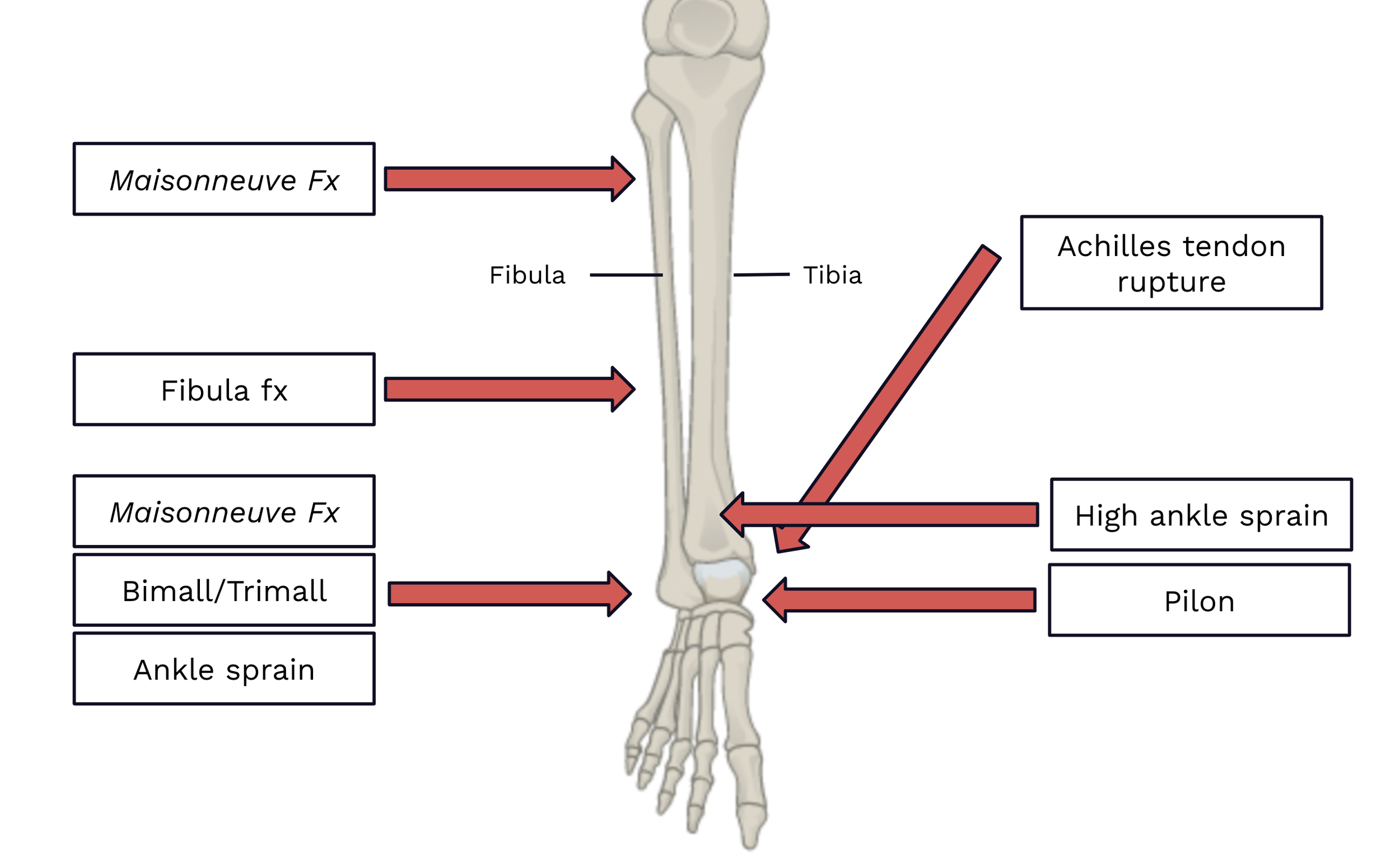
APPROACH TO ANKLE INJURIES
Presentation and Mechanism
Ankle injuries are most commonly caused by abrupt twisting of the ankle. Ankle sprains are most commonly caused by ankle inversion. Ankle fractures may present similarly to an ankle sprain, but are also seen in higher mechanisms of trauma, including MVCs, falls from height, penetrating wounds including GSWs, or other types of direct force to the ankle. Unstable injuries may be obvious at time of presentation, with the patient not being able to bear any weight.
Certain injuries are classically associated with certain mechanisms (e.g., traumatic axial loading with calcaneal fractures, pilons, and vertebral compression fractures) [5]. While knowing the mechanism can provide clues to potential injuries or increase your index of suspicion for certain injuries, mechanisms of injuries can be variable. Respect, but do not rely upon, the mechanism.
Exam
The exam (along with the history) is critical for proper diagnostic evaluation. Use your exam to guide your initial management as well as your imaging decisions and differential diagnosis.
Any significant bleeding should first be controlled in the setting of an open injury
Neurovascular assessment (perfusion, pulses, sensory function, motor function) should be prioritized, regardless of acuity. Abnormal neurovascular findings suggest injury to nearby structures (nerves and blood vessels) from dislocations, bony trauma, or increased compartment pressures.
Gross deformities often suggest fracture or dislocation [Image 2]. Obvious tendon or muscular deformity may be seen with Achilles tendon rupture [Image 3].
Skin tenting or other overlying skin changes are often indicative of impending open fracture or risk of skin necrosis. Significant swelling over a fracture may produce bullae known as “fracture blisters” [Image 4]
Palpate all bones and joints. In general, consider imaging of joints adjacent to an area of bony tenderness and imaging of bones adjacent to areas of joint tenderness.
Range all joints. If active ROM is impaired, perform passive ROM. Impaired ROM can be indicative of dislocation, ligamentous injury, or intra-articular pathology.
Certain provocative tests or inability to bear weight can be clues to instability. If instability is suspected or apparent, avoid having the patient bear weight.
In the setting of a more diffuse traumatic injury, always examine all other bones and joints even if the patient is only complaining of pain at the ankle (their ankle injury may be a distracting injury).
Consider examination and diagnostic evaluation for any injuries that are likely to co-occur (e.g., pilons with fibular fractures, bilateral calcaneal fractures, and vertebral compression fractures [6]).
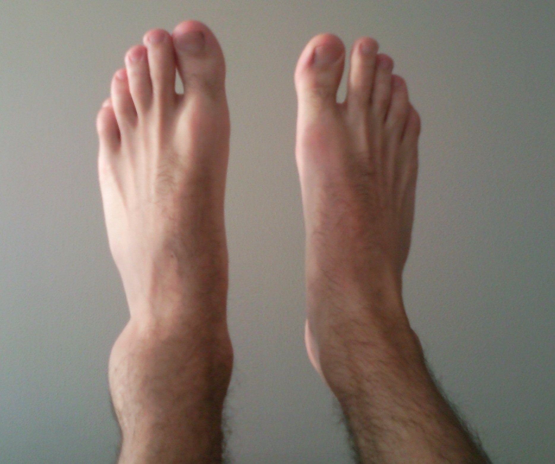
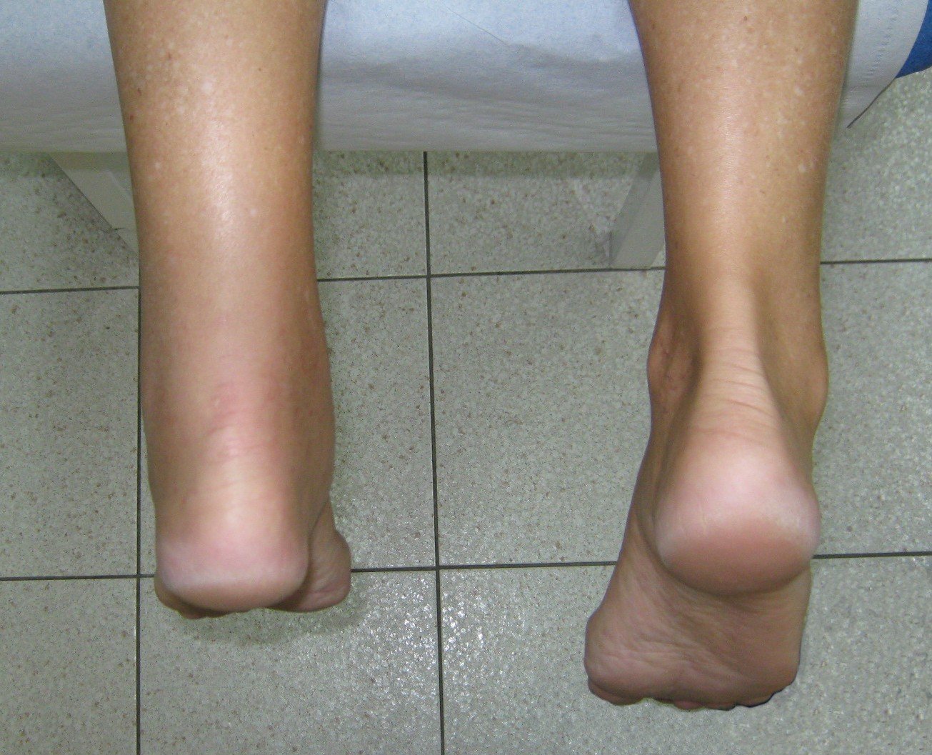
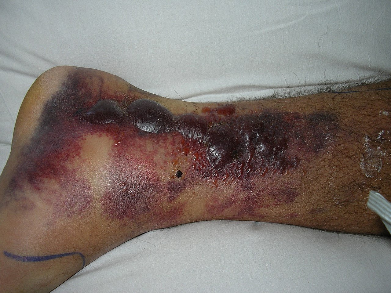
Appropriate imaging
The vast majority of ankle injuries require at least basic radiographs for proper evaluation. Obtain at least 3 views when possible. While the Ottawa Ankle Rules [7] are often referenced and tested on exams, these criteria can miss significant injuries. You should consider full-length tib/fib films in all ankle injuries to rule out a more proximal fracture including a Maisonneuve fracture. Stress films are useful in assessing potential instability by assessing for any widening of the ankle mortise or syndesmosis. Some fractures are often difficult to see on XRs, so consider CT if your index of suspicion is high. Certain injuries require CTs for further characterization or surgical planning. MRI can be used in place of CT. Point-of-care ultrasound (POCUS) can evaluate for Achilles tendon rupture with a high sensitivity (96-100%) and specificity (83-100%) [8,9].
Management
Immediate management:
Control significant bleeding (e.g., direct pressure, tourniquet)
Address any impairments in neurovascular status (e.g., role of consulting services, emergent closed reduction, need for vascular imaging).
In the ED:
Pain control, including consideration for nerve blocks in certain injuries
Wound management (irrigation and debridement, possible closure, consideration of Abx, tetanus immunization)
Closed reduction and/or splinting (make sure to obtain repeat XRs after)
Determine if injury is stable vs. unstable (inherent to injury or as evidenced on stress films or advanced imaging)
Determine if injury is operative vs. non-operative
Determine if Orthopedics (or other specialist) consultation is needed
Unstable or surgical injury (or if this is suspected)
Uncertainty in diagnosis
Uncertainty in management (e.g., weight-bearing status, operative vs. non-operative)
If necessary/helpful to ensure adequate follow-up
At time of discharge:
Counsel on expected course of injury, supportive care (e.g., RICE), and symptomatic management (e.g., NSAIDs, Tylenol, opioid pain medication)
Counsel on ER return precautions, complications that are more likely in certain injuries (e.g., DVT, compartment syndrome), and consider prophylactic measures as appropriate
Brace, other cast, walking boot, immobilizer, etc.
Crutches and crutch training
Give clear instructions on weight-bearing status and use of any braces or walking boots given
Ensure there is a clear follow-up plan and provide any necessary referrals
Who they should see (e.g., primary care physician, Orthopedics, other specialist)
Whether they need to be seen definitely or only if needed, and when
Confirm correct patient contact information in EMR
Open fractures and injuries
Wounds that are not open fractures should receive irrigation and debridement, closure (if indicated), tetanus vaccination, consideration of antibiotics, and wound care instructions. Orthopedics should be consulted for any wounds near joints with concern of extension into the joint (i.e., traumatic arthrotomy), with a lower threshold to consult if the joint is non-native.
Open fractures and dislocations are generally managed with bleeding control, close assessment of neurovascular status, antibiotics with routine gram-positive coverage (Ancef; clindamycin if penicillin allergy), tetanus vaccination, and early irrigation and debridement in the ER with low pressure normal saline [10]. Open fractures can be categorized according to the Gustilo-Anderson classification system [11], though this has been critiqued for poor interrater reliability. Smaller wounds may not require antibiotics, but if in doubt, one dose of Ancef in the ED is unlikely to cause significant harm. Larger wounds (Gustilo-Anderson Type III) should also receive gram-negative microbial coverage. Wounds with potential soil or fecal contamination should receive high-dose penicillin [10]. The management of open fractures is further discussed in these TamingTheSRU posts:
Table 1a Common Ankle Injuries and their management considerations
Table 1B Common Ankle Injuries and their management considerations
References
Bergh C, Wennergren D, Möller M, Brisby H. Fracture incidence in adults in relation to age and gender: A study of 27,169 fractures in the Swedish Fracture Register in a well-defined catchment area. PLoS ONE. 2020.15(12): e0244291. doi.org/10.1371/journal.pone.0244291
Rosenbaum AJ et al. Musculoskeletal health literacy in patients with foot and ankle injuries: A cross-sectional survey of comprehension. Foot & Ankle Specialist. 2016. 9(1):31-36. doi: 10.1177/1938640015593078
Weiss AJ, Jiang J. Most frequent reasons for emergency department visits, 2018. Agency for Healthcare Research and Quality. Statistical brief #286. 2021. http://www.hcup-us.ahrq.gov/reports/statbriefs/sb286-ED-Frequent-Conditions-2018.pdf. Accessed March 5, 2023.
Belatti DA, Phisitkul P. Economic burden of foot and ankle surgery in the US Medicare population. Foot & Ankle Int. 2014,35(4):334-340. doi: 10.1177/1071100713519777
Forsthoefel C. Calcaneus fractures. [Updated 2022 Nov 23]. In: OrthoBullets [Internet]. Available from: https://www.orthobullets.com/trauma/1051/calcaneus-fractures
Fares A, Szatkowski J. Tibial plafond fractures. [Updated 2023 Apr 22]. In: OrthoBullets [Internet]. Available from: https://www.orthobullets.com/trauma/1046/tibial-plafond-fractures
Pires R, Pereira A, Abreu-E-Silva G, Labronici P, Figueiredo L, Godoy-Santos A, Kfuri M. Ottawa ankle rules and subjective surgeon perception to evaluate radiograph necessity following foot and ankle sprain. Ann Med Health Sci Res. 2014 May;4(3):432-5. doi: 10.4103/2141-9248.133473. PMID: 24971221; PMCID: PMC4071746.
Hartgerink P, Fessell DP, Jacobson JA, van Holsbeeck MT. Full versus partial-thickness Achilles tendon tears: sonographic accuracy and characterization in 26 cases with surgical correlation. Radiology. 2001;220:406-12.
Paavola M, Paakkala T, Kannus P, Jarvinen M. Ultrasonography in the differential diagnosis of Achilles tendon injuries and related disorders. A comparison between pre-operative ultrasonography and surgical findings. Acta Radiol. 1998;39(6):612-9.
Garner MR, Sethuraman SA, Schade MA, Boateng H. Antibiotic Prophylaxis in Open Fractures: Evidence, Evolving Issues, and Recommendations. J Am Acad Orthop Surg. 2020 Apr 15;28(8):309-315. doi: 10.5435/JAAOS-D-18-00193. PMID: 31851021.
Kim PH, Leopold SS. Gustilo-Anderson Classification. Clin Orthop Relat Res. 2012 Nov; 470(11):3270-3274. doi: 10.1007/s11999-012-2376-6 PMID: 22569719
Macknet D, Weatherford B. Ankle sprain. [Updated 2023 Feb 26]. In: OrthoBullets. Available from: https://www.orthobullets.com/foot-and-ankle/7028/ankle-sprain
Karadesh M. High ankle sprain & syndesmosis injury. [Updated 2022 Apr 7]. In: OrthoBullets. Available from: https://www.orthobullets.com/foot-and-ankle/7029/high-ankle-sprain-and-syndesmosis-injury
Wong PK, Hanna TN, Shuaib W, Sanders SM, Khosa F. What's in a name? Lower extremity fracture eponyms (Part 2). Int J Emerg Med. 2015 Dec;8(1):76. doi: 10.1186/s12245-015-0076-1. Epub 2015 Jul 25. PMID: 26223985; PMCID: PMC4512960.
Gomez A, Cadogan M. Danis-Weber classification. [Updated 2022 Mar 16]. In: LITFL - Life in the Fast Lane [Internet]. Available from: https://litfl.com/danis-weber-classification/
Taylor BC, Tarazona D. Ankle fractures. [Updated 2023 Apr 11]. In: OrthoBullets [Internet]. Available from: https://www.orthobullets.com/trauma/1047/ankle-fractures
Koujan K, Saber AY. Bimalleolar Ankle Fractures. [Updated 2023 Feb 11]. In: StatPearls [Internet]. Treasure Island (FL): StatPearls Publishing; 2023 Jan-. Available from: https://www.ncbi.nlm.nih.gov/books/NBK562254/
Authorship
Written by: Isabel Lott, MD, PGY-1, University of Cincinnati Department of Emergency Medicine
Peer Review by Bret Betz, MD, Associate Professor, University of Cincinnati Department of Emergency Medicine
Editing and Posting by Jeffery Hill, MD MEd, Associate Professor, University of Cincinnati Department of Emergency Medicine
Cite As
Lott, I., Betz, B., Hill, J. (May 22, 2023) Twisting and Turning - Ankle Injuries in the ED. TamingtheSRU. https://www.tamingthesru.com/blog/diagnostics/ankle-injuries-in-the-ed

The CLOVERS Trial
Sepsis, including severe sepsis and septic shock, is a frequently encountered condition in the emergency department and carries a high mortality rate. One of the primary pathophysiologic mechanisms involves complex cascade of host dysregulation in response to an infectious stimulus (Evans, Rhodes et al. 2021, Jarczak, Kluge et al. 2021). Recent meta-analyses and systematic reviews evaluating mortality in patients with septic shock reported mortality as high as 35% and 38% at 30 and 90 days, respectively (Vincent, Jones et al. 2019, Bauer, Gerlach et al. 2020). Despite the complexity and heterogeneity of patients with sepsis, there have been few interventions which have been demonstrated to decrease mortality: early antimicrobial and fluid administration (Levy, Evans et al. 2018, Kuttab, Lykins et al. 2019, Evans, Rhodes et al. 2021, Im, Kang et al. 2022), ideally with antibiotics administered within one hour of sepsis recognition by the treating provider (Evans, Rhodes et al. 2021). Each subsequent one-hour delay in antimicrobial administration increases mortality by 35% in patients with septic shock (Im, Kang et al. 2022).
National Heart, Lung, and Blood Institute Prevention and Early Treatment of Acute Lung Injury Clinical Trials Network, Shapiro NI, Douglas IS, et al. Early Restrictive or Liberal Fluid Management for Sepsis-Induced Hypotension. N Engl J Med. 2023;388(6):499-510. doi:10.1056/NEJMoa2212663
BACKGROUND
Sepsis, including severe sepsis and septic shock, is a frequently encountered condition in the emergency department and carries a high mortality rate. One of the primary pathophysiologic mechanisms involves complex cascade of host dysregulation in response to an infectious stimulus (Evans, Rhodes et al. 2021, Jarczak, Kluge et al. 2021). Recent meta-analyses and systematic reviews evaluating mortality in patients with septic shock reported mortality as high as 35% and 38% at 30 and 90 days, respectively (Vincent, Jones et al. 2019, Bauer, Gerlach et al. 2020). Despite the complexity and heterogeneity of patients with sepsis, there have been few interventions which have been demonstrated to decrease mortality: early antimicrobial and fluid administration (Levy, Evans et al. 2018, Kuttab, Lykins et al. 2019, Evans, Rhodes et al. 2021, Im, Kang et al. 2022), ideally with antibiotics administered within one hour of sepsis recognition by the treating provider (Evans, Rhodes et al. 2021). Each subsequent one-hour delay in antimicrobial administration increases mortality by 35% in patients with septic shock (Im, Kang et al. 2022).
The SSC guidelines recommend administering at least 30mL/kg of crystalloid within a three hour period for patients with sepsis-associated hypoperfusion and septic shock (Evans, Rhodes et al. 2021), although this is based on low-quality evidence rather than high-quality randomized trials and has remained a controversial aspect of treatment of patients with sepsis given the potential harms of large-volume fluid administration. In fact, specifically within the SSC guidelines the evidence is described verbatim as “weak recommendation, low-quality evidence” (Evans, Rhodes et al. 2021).
Several studies have begun exploring the potential link between large-volume fluid administration and mortality and end-organ harm in patients with sepsis and septic shock. Respiratory failure and more severe acute kidney injury were demonstrated to be more common in patients who received higher volumes of intravenous fluid (Andrews, Muchemwa et al. 2014, Hjortrup, Haase et al. 2016, Andrews, Semler et al. 2017, Silversides, Major et al. 2017). A systematic review and meta-analysis of several cohort studies demonstrated 70% increased mortality (pooled RR: 1.70; CI: 1.20, 2.41; P = .003) with high-volume fluid administration in patients with severe sepsis and septic shock, and significantly lower mortality with low-volume fluid administration in the first 24h of treatment (P=0.02) (Tigabu, Davari et al. 2018). This was corroborated by another systematic review which demonstrated decreased mortality in restrictive compared to liberal fluid administration strategies (24.7% vs 33.2%; OR, 0.42; 95% CI 0.32-0.55; P < 0.0001) (Malbrain, Marik et al. 2014).
The ANDROMEDA-SHOCK trial published in 2019 demonstrated that only one-quarter (25%) of patients were fluid-responsive at baseline, as determined most frequently by velocity time integral or passive leg raise methods (Hernández, Ospina-Tascón et al. 2019). In 2022, the CLASSIC trial was published which provided one investigational angle to this important question. The CLASSIC trial is a randomized controlled trial conducted in the ICU setting which compared restrictive versus standard fluid strategies with a primary outcome of death by 90 days in patients with septic shock. This trial involved employing a restrictive versus standard strategy with the restrictive fluid strategy permitting fluid boluses only when the patient met one of several criteria, whereas the standard fluid strategy had no limit to fluid quantity administered. Mortality at 90 days was equivocal between groups (42.3% vs 42.1%), as were days alive outside of the hospital and days without life support (Meyhoff, Hjortrup et al. 2022).
The CLASSIC trial nicely set up for the introduction and publishing of the primary study of focus of this post:
The CLOVERS 🍀 Trial
(National Heart, Lung, and Blood Institute Prevention and Early Treatment of Acute Lung Injury Clinical Trials Network, Shapiro NI, Douglas IS, et al. Early Restrictive or Liberal Fluid Management for Sepsis-Induced Hypotension. N Engl J Med. 2023;388(6):499-510. doi:10.1056/NEJMoa2212663)
METHODS
Setting
The CLOVERS trial is a multi-center, unblinded, superiority randomized controlled trial which functioned as part of the PETAL (Prevention and Early Treatment of Acute Lung Injury Network) network. Sixty centers participated during the years 2018-2022.
Inclusion criteria
Adults (18 years and older) with “suspected or confirmed infection” with sepsis-induced hypotension, which was defined as SBP less than 100 after 1L or greater of crystalloid administration, were included.
Exclusion criteria
Patients were excluded if:
It had been greater than four hours since meeting criteria for sepsis-induced hypotension (SBP <100 after 1L≤ crystalloid administered).
Greater than 24h since presentation to the hospital
Having received greater than 3L intravenous fluids – including prehospital volume administration
Evidence of volume overload (importantly, did not exclude patients carrying a diagnosis of heart failure or end-stage renal disease by diagnosis alone)
Evidence of severe volume depletion for other causes (ex. elderly patient with HHS found down with severe hypovolemic shock, hypernatremia, etc.)
Randomization
This was accomplished via a Web-based centralized system with site-based stratification.
Intervention
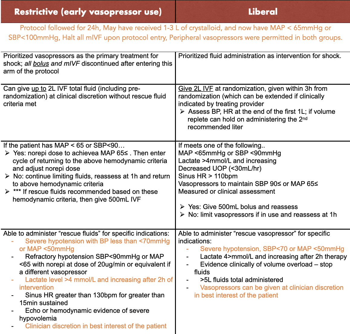
Protocol
Adherence was amended in October of 2019 to limit administered fluid bolus to 1L if vital signs had stabilized (noted in chart above). The study was stopped at the second interim analysis due to futility – rather than harm from the DSMB. However, protocol adherence was monitored for the first 300 patients and during the remainder of the trial.
Study power
They assumed at baseline mortality of 15% and the study was powered to detect an absolute difference of 4.5% in the restrictive fluid group. 2,320 patients were needed to have 90% power at a significance level of 0.05.
RESULTS
A total of 1,563 patients were enrolled with 782 assigned to the restrictive and 781 assigned to the liberal fluid groups. There were similar baseline characteristics and the groups were well-matched. Before randomization, both groups received similar fluid volumes (median 2050mL for both) and similar percentages of vasopressor use (21% in restrictive, 18% in liberal).
Fluid volumes between groups at the six-hour mark differed – with a median of 500mL and 2300mL in the restrictive and liberal fluid groups respectively. At the 24h mark, the median volumes were 1267mL and 3400mL (restrictive, liberal) and mean difference of -2134mL. 59% of patients in the restrictive and 37% in liberal fluid groups received vasopressors. Lactated ringer’s solution was the most frequently chosen fluid type.
Adherence
Protocol adherence was remarkably high – 97% vs 96% in the restrictive and liberal groups and sustained throughout the duration of the trial.
Post-hoc analysis
There were some post-hoc analyses that were conducted including more patients in the restrictive group were admitted to the intensive care unit versus the liberal group (67.3% versus 59.2%). No effects stratified by site were detected.
Primary outcome
There was no significant difference in the primary outcome, death before discharge home by 90 days between groups (14.0% versus 14.9% in restrictive versus liberal; estimated difference -0.9 percentage points; 95% CI -4.4 to 2.6; P=0.61). Subgroup analyses which were prespecified such as chronic heart failure, ESRD, pneumonia as cause, and receipt of vasopressors or SBP<90mmHg at randomization were conducted and reported in detail in the primary literature, but demonstrated interestingly a mortality difference of -20.2% for patients with ESRD in the restrictive group (95% CI; -41.9 to 1.5).
Safety outcomes
Serious adverse event occurrences were similar in both groups. For both volume overload and pulmonary edema, each occurred only 3 times in the liberal group versus 0 in the restrictive group. There were only three possible events of vasopressor extravasation, among 500 patients, who did receive vasopressors, all of which resolved without intervention and left no residual effects on the patient.
DISCUSSION
This was a well-conducted randomize controlled trial evaluating an important clinical question, and importantly, was conducted in an emergency department setting and has a significant strength and addition to the literature in that this is a prospective trial. It would be difficult to perform a blinded trial of similar nature – although this does provide for some limitation on potential biases. Unfortunately, it was ultimately underpowered and enrollment was ceased early secondary to futility as determined by the DSMB. Additionally, some other studies have identified a higher mortality than prespecified in the methods section (15%) – closer to ~35% - and raises the question of similarity to some patient populations, which may be much more critically ill at baseline. The protocol was well-designed, however also had to weigh the ml/kg versus absolute fluid volume for standardization; would 30mL/kg have been a better initial comparator group rather than absolute untailored fluid amounts, based on the Surviving Sepsis Campaign guidelines?
A 90-day timeline outcome was used similarly as an outcome to the CLASSIC trial; this is a patient-centered outcome, and corroborates the emergency department counterpiece to the ICU-based CLASSIC trial suggesting no difference in 90-day discharge home and 90-day mortality between the two trials. However – is this the best endpoint for such a trial? 90 days accounts for significant post-treatment (given 24h protocol) variability which is influenced by a grand multitude of factors.
Both restrictive and more liberal protocols for the treatment of septic shock appear to be viable initial resuscitation strategies, and importantly, safety was maintained in both – including for the peripheral administration of vasopressors which is a key point for emergency providers who may have limitations on time and bandwidth in caring for many patients simultaneously in the emergency department and could limit performing unnecessary central lines and invasive procedures on patients. It would be interesting to stratify the doses of vasopressor administration to the few potential adverse events (all of which resolved), or the location of the peripheral line, given the questions raised regarding safety and patency of ultrasound-guided IVs versus non-ultrasound guided-IVs.
Specific patient populations were excluded, of note, such as the septic, already-volume-overloaded or severely hypovolemic patient – these will require careful, separate consideration in the tailored, specialized treatment of septic shock. There was also an advantage of using MAP as an endpoint, which is a significant advantage as this is a standard resuscitation and hemodynamic end point in routine clinical care of patients in shock. Although protocol adherence was high, one challenge that is pervasive between studies of similar hypothesis regarding fluid management, is heterogeneity of the treatment effects. There was significant crossover in baseline vasopressor administration, for example – and although this is appropriate for patient safety and for clinical care flexibility, it does somewhat cloud the ultimate interpretation and treatment effects.
Overall, both restrictive and liberal fluid strategies in the initial resuscitation of patients with septic shock, when applied to the appropriate patient populations, are viable strategies for care based on the provided endpoints and subgroup and post-hoc analyses. This study raised some interesting potential questions and hypotheses for future studies based on the subgroup analyses, and demonstrated excellent safety for the administration of peripheral vasopressors.
REFERENCES
Andrews, B., L. Muchemwa, P. Kelly, S. Lakhi, D. C. Heimburger and G. R. Bernard (2014). "Simplified severe sepsis protocol: a randomized controlled trial of modified early goal-directed therapy in Zambia." Crit Care Med 42(11): 2315-2324.
Andrews, B., M. W. Semler, L. Muchemwa, P. Kelly, S. Lakhi, D. C. Heimburger, C. Mabula, M. Bwalya and G. R. Bernard (2017). "Effect of an Early Resuscitation Protocol on In-hospital Mortality Among Adults With Sepsis and Hypotension: A Randomized Clinical Trial." Jama 318(13): 1233-1240.
Bauer, M., H. Gerlach, T. Vogelmann, F. Preissing, J. Stiefel and D. Adam (2020). "Mortality in sepsis and septic shock in Europe, North America and Australia between 2009 and 2019- results from a systematic review and meta-analysis."Crit Care 24(1): 239.
Evans, L., A. Rhodes, W. Alhazzani, M. Antonelli, C. M. Coopersmith, C. French, F. R. Machado, L. McIntyre, M. Ostermann, H. C. Prescott, C. Schorr, S. Simpson, W. J. Wiersinga, F. Alshamsi, D. C. Angus, Y. Arabi, L. Azevedo, R. Beale, G. Beilman, E. Belley-Cote, L. Burry, M. Cecconi, J. Centofanti, A. Coz Yataco, J. De Waele, R. P. Dellinger, K. Doi, B. Du, E. Estenssoro, R. Ferrer, C. Gomersall, C. Hodgson, M. H. Moller, T. Iwashyna, S. Jacob, R. Kleinpell, M. Klompas, Y. Koh, A. Kumar, A. Kwizera, S. Lobo, H. Masur, S. McGloughlin, S. Mehta, Y. Mehta, M. Mer, M. Nunnally, S. Oczkowski, T. Osborn, E. Papathanassoglou, A. Perner, M. Puskarich, J. Roberts, W. Schweickert, M. Seckel, J. Sevransky, C. L. Sprung, T. Welte, J. Zimmerman and M. Levy (2021). "Surviving sepsis campaign: international guidelines for management of sepsis and septic shock 2021." Intensive Care Med 47(11): 1181-1247.
Hernández, G., G. A. Ospina-Tascón, L. P. Damiani, E. Estenssoro, A. Dubin, J. Hurtado, G. Friedman, R. Castro, L. Alegría, J. L. Teboul, M. Cecconi, G. Ferri, M. Jibaja, R. Pairumani, P. Fernández, D. Barahona, V. Granda-Luna, A. B. Cavalcanti, J. Bakker, G. Hernández, G. Ospina-Tascón, L. Petri Damiani, E. Estenssoro, A. Dubin, J. Hurtado, G. Friedman, R. Castro, L. Alegría, J. L. Teboul, M. Cecconi, M. Cecconi, G. Ferri, M. Jibaja, R. Pairumani, P. Fernández, D. Barahona, A. B. Cavalcanti, J. Bakker, G. Hernández, L. Alegría, G. Ferri, N. Rodriguez, P. Holger, N. Soto, M. Pozo, J. Bakker, D. Cook, J. L. Vincent, A. Rhodes, B. P. Kavanagh, P. Dellinger, W. Rietdijk, D. Carpio, N. Pavéz, E. Henriquez, S. Bravo, E. D. Valenzuela, M. Vera, J. Dreyse, V. Oviedo, M. A. Cid, M. Larroulet, E. Petruska, C. Sarabia, D. Gallardo, J. E. Sanchez, H. González, J. M. Arancibia, A. Muñoz, G. Ramirez, F. Aravena, A. Aquevedo, F. Zambrano, M. Bozinovic, F. Valle, M. Ramirez, V. Rossel, P. Muñoz, C. Ceballos, C. Esveile, C. Carmona, E. Candia, D. Mendoza, A. Sanchez, D. Ponce, D. Ponce, J. Lastra, B. Nahuelpán, F. Fasce, C. Luengo, N. Medel, C. Cortés, L. Campassi, P. Rubatto, N. Horna, M. Furche, J. C. Pendino, L. Bettini, C. Lovesio, M. C. González, J. Rodruguez, H. Canales, F. Caminos, C. Galletti, E. Minoldo, M. J. Aramburu, D. Olmos, N. Nin, J. Tenzi, C. Quiroga, P. Lacuesta, A. Gaudín, R. Pais, A. Silvestre, G. Olivera, G. Rieppi, D. Berrutti, M. Ochoa, P. Cobos, F. Vintimilla, V. Ramirez, M. Tobar, F. García, F. Picoita, N. Remache, V. Granda, F. Paredes, E. Barzallo, P. Garcés, F. Guerrero, S. Salazar, G. Torres, C. Tana, J. Calahorrano, F. Solis, P. Torres, L. Herrera, A. Ornes, V. Peréz, G. Delgado, A. López, E. Espinosa, J. Moreira, B. Salcedo, I. Villacres, J. Suing, M. Lopez, L. Gomez, G. Toctaquiza, M. Cadena Zapata, M. A. Orazabal, R. Pardo Espejo, J. Jimenez, A. Calderón, G. Paredes, J. L. Barberán, T. Moya, H. Atehortua, R. Sabogal, G. Ortiz, A. Lara, F. Sanchez, A. Hernán Portilla, H. Dávila, J. A. Mora, L. E. Calderón, I. Alvarez, E. Escobar, A. Bejarano, L. A. Bustamante and J. L. Aldana (2019). "Effect of a Resuscitation Strategy Targeting Peripheral Perfusion Status vs Serum Lactate Levels on 28-Day Mortality Among Patients With Septic Shock: The ANDROMEDA-SHOCK Randomized Clinical Trial." Jama 321(7): 654-664.
Hjortrup, P. B., N. Haase, H. Bundgaard, S. L. Thomsen, R. Winding, V. Pettilä, A. Aaen, D. Lodahl, R. E. Berthelsen, H. Christensen, M. B. Madsen, P. Winkel, J. Wetterslev and A. Perner (2016). "Restricting volumes of resuscitation fluid in adults with septic shock after initial management: the CLASSIC randomised, parallel-group, multicentre feasibility trial." Intensive Care Med 42(11): 1695-1705.
Im, Y., D. Kang, R. E. Ko, Y. J. Lee, S. Y. Lim, S. Park, S. J. Na, C. R. Chung, M. H. Park, D. K. Oh, C. M. Lim, G. Y. Suh and i. Korean Sepsis Alliance (2022). "Time-to-antibiotics and clinical outcomes in patients with sepsis and septic shock: a prospective nationwide multicenter cohort study." Crit Care 26(1): 19.
Jarczak, D., S. Kluge and A. Nierhaus (2021). "Sepsis-Pathophysiology and Therapeutic Concepts." Front Med (Lausanne) 8: 628302.
Kuttab, H. I., J. D. Lykins, M. D. Hughes, K. Wroblewski, E. P. Keast, O. Kukoyi, J. A. Kopec, S. Hall and M. A. Ward (2019). "Evaluation and Predictors of Fluid Resuscitation in Patients With Severe Sepsis and Septic Shock." Crit Care Med 47(11): 1582-1590.
Levy, M. M., L. E. Evans and A. Rhodes (2018). "The Surviving Sepsis Campaign Bundle: 2018 update." Intensive Care Med 44(6): 925-928.
Malbrain, M. L., P. E. Marik, I. Witters, C. Cordemans, A. W. Kirkpatrick, D. J. Roberts and N. Van Regenmortel (2014). "Fluid overload, de-resuscitation, and outcomes in critically ill or injured patients: a systematic review with suggestions for clinical practice." Anaesthesiol Intensive Ther 46(5): 361-380.
Meyhoff, T. S., P. B. Hjortrup, J. Wetterslev, P. Sivapalan, J. H. Laake, M. Cronhjort, S. M. Jakob, M. Cecconi, M. Nalos, M. Ostermann, M. Malbrain, V. Pettila, M. H. Moller, M. N. Kjaer, T. Lange, C. Overgaard-Steensen, B. A. Brand, M. Winther-Olesen, J. O. White, L. Quist, B. Westergaard, A. B. Jonsson, C. J. S. Hjortso, N. Meier, T. S. Jensen, J. Engstrom, L. Nebrich, N. C. Andersen-Ranberg, J. V. Jensen, N. A. Joseph, L. M. Poulsen, L. S. Herlov, C. G. Solling, S. K. Pedersen, K. K. Knudsen, T. S. Straarup, M. L. Vang, H. Bundgaard, B. S. Rasmussen, S. R. Aagaard, T. Hildebrandt, L. Russell, M. H. Bestle, M. Schonemann-Lund, A. C. Brochner, C. F. Elvander, S. K. L. Hoffmann, M. L. Rasmussen, Y. K. Martin, F. F. Friberg, H. Seter, T. N. Aslam, S. Adnoy, P. Seidel, K. Strand, B. Johnstad, E. Joelsson-Alm, J. Christensen, C. Ahlstedt, C. A. Pfortmueller, M. Siegemund, M. Greco, J. Radej, M. Kriz, D. W. Gould, K. M. Rowan, P. R. Mouncey, A. Perner and C. T. Group (2022). "Restriction of Intravenous Fluid in ICU Patients with Septic Shock." N Engl J Med 386(26): 2459-2470.
Silversides, J. A., E. Major, A. J. Ferguson, E. E. Mann, D. F. McAuley, J. C. Marshall, B. Blackwood and E. Fan (2017). "Conservative fluid management or deresuscitation for patients with sepsis or acute respiratory distress syndrome following the resuscitation phase of critical illness: a systematic review and meta-analysis." Intensive Care Med 43(2): 155-170.
Tigabu, B. M., M. Davari, A. Kebriaeezadeh and M. Mojtahedzadeh (2018). "Fluid volume, fluid balance and patient outcome in severe sepsis and septic shock: A systematic review." J Crit Care 48: 153-159.
Vincent, J. L., G. Jones, S. David, E. Olariu and K. K. Cadwell (2019). "Frequency and mortality of septic shock in Europe and North America: a systematic review and meta-analysis." Crit Care 23(1): 196.
Authorship
Written by Lauren Gillespie, MD, PGY-3, University of Cincinnati Department of Emergency Medicine
Peer Review, Editing, Audio Editing, Posting by Jeffery Hill, MD MEd
Cite As
Gillespie, L., Hill, J. (May 5, 2023) The CLOVERS Trial. TamingtheSRU. https://www.tamingthesru.com/blog/journal-club/the-clovers-trial

Air Care Series: Epoprostenol/VELETRI: a Skybridge to Somewhere?
Ever have that critical pneumonia ARDS patient that just cannot be safely transported without a temporizing (or longer) fix? Join Dr. Ferreri on a case study and deep dive into the physiology behind Epoprostenol use in Transport Medicine.
CLINICAL CASE:
History of Present Illness:
An elderly female initially presented to an outside hospital with a variety of respiratory symptoms and was subsequently diagnosed with COVID-19 pneumonia. During her hospitalization, her respiratory status gradually deteriorated and five days into her hospitalization, she unfortunately required endotracheal intubation. Despite mechanical ventilation, she developed refractory hypoxia. Initial attempts were made to increase the PEEP from 5 to 15cm H2O, to little avail. On the eighth day of admission, the decision was then made to paralyze and prone the patient. A follow-up chest x-ray at that time revealed pneumomediastinum, and she was started on broad-spectrum antibiotics. Ultimately, on the cusp of deterioration, the patient was deemed most appropriate for transfer to a medical intensive care unit (MICU) at a large academic hospital via helicopter emergency medical services (HEMS).
On arrival of the UC Air Care team, the patient was found to be intubated, sedated, and paralyzed in the prone position. Ventilator settings were ARVC with an FiO2 80%, RR 24bpm, Vt 450cc, and a PEEP 12cm H20.
While preparing for transport, the patient was placed in the supine position, leading to an acute desaturation event to 85%. The HEMS team did not feel that air transport in the prone position would be a safe option and needed other methods to improve the patient’s oxygenation. While still in the supine position, inhaled epoprostenol (aka VELETRI) was initiated and the set FiO2 was increased to 100%. Much to the relief of the team, these interventions rapidly improved her SpO2 readings to 94-96%. While preparing the patient and equipment for loading into the helicopter, she became hypotensive and required 10mcg of push-dose epinephrine.
In flight, a rapid blood gas analysis revealed a prominent respiratory acidosis with hypercapnia, prompting the set respiratory rate to be raised. Meanwhile, as the SpO2 remained above 90%, FiO2 was gradually weaned down. She was noted to be persistently hypotensive, with systolic blood pressures in the 90’s, and the norepinephrine drip rate was titrated to 20 mcg/min. She was successfully unloaded from the helicopter and her care was relinquished to the MICU team eagerly awaiting her arrival.
Hospital Course:
On arrival to the MICU, a chest x-ray was performed and revealed a moderate left-sided pneumothorax associated with pneumomediastinum and diffuse airspace disease. A chest tube was placed with improvement in her respiratory status, allowing ventilator settings to be gradually liberated. Additionally, epoprostenol was weaned and then discontinued 24 hours after her initial arrival. During the admission, she completed a course of broad-spectrum antibiotics and as the underlying shock state improved, she was also weaned off all vasopressor support. She was noted to have improvement in her underlying respiratory status as well and was extubated approximately three weeks later.
With time, the patient serially progressed down the various levels of medical care. On hospital day 38, she was successfully discharged to a skilled nursing facility, requiring just a nasal cannula for supplemental oxygen support, as well as a prescription for apixaban for new-onset atrial fibrillation.
DISCUSSION:
This case highlights changes that have been made in the arena of critical care transport, specifically UC Air Care transport, during the COVID-19 pandemic. Often, patients with refractory hypoxia or pulmonary artery hypertension previously have had epoprostenol started prior to initiating transportation. In the wake of the pandemic, we now carry epoprostenol on all flights for use on patients with severe refractory hypoxemia, right heart failure, and/or pulmonary hypertension. It is important to understand how this drug is delivered, its basic pharmacology, and ultimately how it can be leveraged to improve our patients’ care enroute to a definitive treatment facility.
INHALDED PULMONARY VASODILATORS: EPOPROSTENOL
Epoprostenol and Prostacyclin Analogs:

Epoprostenol is a prostacyclin analogue that acts at the prostaglandin I receptor, specifically the prostaglandin I2 receptor (PGI2). This is a G protein-coupled receptor (GPCR) found on platelets, smooth muscles, and immune cell membranes. Once bound, GPCR activation leads to increased release of cyclic AMP (cAMP). Elevation in cAMP leads to inhibition of platelet aggregation, smooth muscle relaxation, and decreased inflammatory cell proliferation (1,2). Smooth muscle relaxation occurs through the inhibition of endothelin release and increased production of nitric oxide (1). When epoprostenol is used via the inhaled route, it causes targeted effects to areas of the lung receiving sufficient ventilation. This targeted pulmonary vasodilation is thought to improve underlying ventilation-perfusion mismatch, leading to increased blood flow to already well-ventilated lung tissue (3). Oxygenation is further improved through decreased pulmonary vascular resistance (PVR) and pulmonary mean arterial pressure (PMAP) (4). Since epoprostenol is often administered in the critical care setting via inhalation, its systemic effects are thought to be limited. However, inhalational administration of epoprostenol requires certain equipment, namely a drug delivery device such as an Aerogen nebulizer (5).
Inhaled prostacyclin, and prostacyclin analogs, are commonly used in the management of acute respiratory distress syndrome (ARDS) or for combatting acute right-sided heart failure. Their actions likely stretch beyond just the vasodilatory properties mentioned above. For instance, agonism on the PGI2 receptors leads to reduced leukocyte adhesion, as well as platelet de-aggregation and antithrombotic effects (6). This likely tapers the large-scale inflammatory response of these disease processes, especially ARDS.

Despite the physiologic advantages associated with the use of prostacyclin, there is a theoretical risk of diffuse pulmonary vasodilation and worsening V/Q mismatching in patients with significant ARDS (7). Further, though aerosolized prostacyclin and their analogs have the advantage of acting primarily on the pulmonary vasculature, systemic absorption can occur as well, leading to systemic hypotension, bleeding, flushing, headache, nausea, vomiting and chest pain (4,8).
Pharmacokinetics:
Epoprostenol is rapidly hydrolyzed in the blood and therefore has a half-life between 2 and 5 minutes (9,10). It is most often delivered via a nebulizer directly into the ventilator circuit of an intubated patient, but it may also be administered via high flow nasal canula and/or CPAP/BiPAP (10). Though uncommon in the critical care setting, it can be administered intravenously as well. Inhalation doses typically range between 0-50 ng/kg/min; however, in the management of critically ill patients, treatment is often started at the maximum dose and then weaned as tolerated (10). Please see the associated image for epoprostenol dosing.
CLINICAL UTILITY OF EPOPROSTENOL:
Acute Respiratory Distress Syndrome
The patient presented in this case was diagnosed with COVID-19 pneumonia complicated by Acute Respiratory Distress Syndrome (ARDS). ARDS is caused by direct and indirect insult to the lungs, which leads to a cascade of inflammatory processes hindering the alveoli and results in various degrees of impaired oxygenation. It is graded from mild to severe, based on the PaO2 to FiO2 ratio (P/F ratio) and can also be formally graded using the Berlin Criteria (11). The cause of death in patients with ARDS is often multisystem organ failure and/or refractory hypoxemia.
ARDS also leads to inadequate ventilation to a high proportion of damaged parenchymal lung tissue. This occurs from the direct injury to the alveolar tissue by surrounding interstitial edema, making it difficult for effective gas exchange to occur despite ventilation. This impaired perfusion at the level of the alveoli is caused by hypoxia, microthrombi, and direct injury to the tissue. This cumulatively leads to pulmonary vasoconstriction and increases the afterload faced by the right side of the heart. ARDS contributes to two main issues, hypoxemia and increased pulmonary vascular resistance, both of which epoprostenol aims to address.
Through the inhaled route, epoprostenol will cause improved V/Q matching through vasodilation of well-ventilated areas of the lung. This will allow improved gas exchange, oxygenation, and in turn improve the P/F ratio.

For a more complete discussion on ARDS, please see this other Air Care Series post on the subject.
Right Ventricular Physiology:
The other prominent effect that epoprostenol has is decreased pulmonary vascular resistance, therefore improving right heart function through a reduction in the perceived RV afterload.
Right ventricular physiology is governed by four key components: 1) systemic venous return, or preload, 2) pulmonary arterial pressure, or right ventricular afterload, 3) right ventricular compliance, and 4) right ventricular free wall / interventricular septum contractility (12). Under normal physiologic circumstances, the right ventricle contracts against a highly compliant and low resistance pulmonary circulation. As a result, the right ventricle is well adapted to changes in volume (12,13). However, the RV experiences a greater change in end-systolic volume as compared to the left ventricle (LV) and is highly sensitive to changes in afterload (12,14,15). Unlike the left ventricle, which receives its blood supply from the coronary arteries during diastole, the right ventricle receives its blood flow during both systole and diastole (16).16 As such, the right ventricle is prone to decreased coronary blood flow and subendocardial ischemia in the setting of increased right ventricular end diastolic pressures, which may exacerbate RV dysfunction.
For a complete discussion of cardiogenic shock and right ventricular failure, please see this other Air Care Series post on the subject.
Pathophysiology of Right Sided Heart Failure:
Though complex and multifactorial, the pathophysiology of acute right-sided heart failure typically involves an abrupt increase in right ventricular afterload (16). Hypoxic vasoconstriction, such as in the setting of ARDS or a pulmonary embolism, is a common underlying and usually reversible cause of an abrupt increase in RV afterload. Furthermore, these rapid changes in pulmonary vascular resistance can lead to significant reduction in RV stroke volume and subsequent RV dilation & tricuspid regurgitation (16). As RV dilation progresses and right-sided filling pressures rise, there is a risk of coronary sinus venous congestion and subsequent RV ischemia, further worsening ventricular function (17,18).
Besides an acute increase in RV afterload, a reduction in RV contractility can also lead to acute right-sided heart failure. This is commonly seen in the setting of an acute myocardial ischemia and can be seen in up to 50% of inferior ST elevation myocardial infarctions (19). Furthermore, structural abnormalities to the myocardium, such as myocarditis, may also contribute to worsened right ventricular contractility. Since the RV does not exist in isolation, its failure goes on to propel hepatic and renal dysfunction via back-up of venous blood. This cumulates to self-enforce a cycle of worsening venous congestion and propagating RV failure (16).
Medical management of right sided heart failure is geared toward optimizing a patient’s preload and right ventricular afterload. Inhaled pulmonary vasodilators, such as epoprostenol, can play a direct role in this process. In patients with acute increases in RV afterload, commonly due to increased pulmonary vasoconstriction from hypoxia or thrombus, inhaled prostacyclin can help reduce RV afterload and ideally temporize RV function.
In the patient case discussed above, inhaled epoprostonel assisted with improved oxygenation and decreased stress on the RV.
Efficacy and Evidence
Epoprostenol is thought to have physiologic benefits in patients with acute right sided heart failure, elevated pulmonary vascular pressures, and ARDS. The use of epoprostenol is often debated, and evidence is lacking regarding the benefits on mortality (7,8). To date there are only a small number of observational studies, and even less randomized control trials.
A recent meta-analysis conducted by Fuller et al. in 2015 included 25 studies, two of which were randomized control trials. Researchers in this study found an increase in the PaO2 to FiO2 ratio in those treated with prostacyclin analogs, as well as a decrease in the mean pulmonary artery pressure in patients. However, in the setting of significant heterogeneity of data as well as reported side effects, the authors concluded that they were unable to reliably attest to the benefit or harm in the use of prostacyclin analogs in patients with ARDS (8).
In a recent Cochrane Review of available literature assessing the efficacy of prostacyclin analog use in the treatment of ARDS, Afshari et al. 2017 conducted a systematic review of two randomized control trials (7,20,21). One of the included studies evaluated the efficacy of inhaled prostacyclin in children (20). The authors found that overall oxygen index was improved significantly among those treated with prostacyclin analog compared to those treated with saline. The other study included those 18 years of age or older and found that there was a non-significant improvement in the PaO2 to FiO2 ratio among those treated with alprostadil compared to those treated with saline (21). Ultimately, the authors of the Cochrane Review concluded that both studies had “very low quality of evidence” and stated that there is insufficient evidence to support the routine use of aerosolized prostacyclin in people with acute respiratory distress syndrome” due to a lack of reliable data on its impact on overall mortality (7). Despite the unclear data on epoprostenol efficacy and its ambiguous effects on overall morbidity & mortality, a recent prospective study found that there may be a potential cost benefit when comparing it to nitric oxide (22).
SUMMARY:
The management of ARDS and right-sided heart failure can be daunting. Fortunately, we have multiple tools to combat refractory hypoxia, elevated pulmonary arterial pressures, and right ventricular failure. Of these, epoprostenol and other prostacyclin analogs have been utilized despite limited data on their actual efficacy. Though promising data has been published endorsing the use of prostacyclin analogs in the treatment of acute right sided heart failure, specifically in the setting of ARDS, there are very few randomized controlled trials providing evidence of its benefit on actual meaningful patient outcomes. Though there may be a cost benefit associated with its use, there is an urgent need for further randomized studies evaluating the use of these aerosolized pulmonary vasodilators and their impact on morbidity &mortality. Though further outcome data is certainly needed, inhaled epoprostenol is just another tool in our arsenal to help stabilize critically-ill patients several thousand feet in the sky, while transferring them to a definite center of care.
AUTHORED BY Josh ferreri, MD
Dr. Ferreri is a PGY-3 in Emergency Medicine at the University of Cincinnati
POSTED BY max kletsel, MD, MS and Christopher Zalesky, MD MSc
Dr. Kletsel is a PGY-3 in Emergency Medicine at the University of Cincinnati
Dr. Zalesky is a PGY-4 in Emergency Medicine at the University of Cincinnati
FACULTY EDITOR william knight, MD
Dr. Knight is a Professor of Emergency Medicine and Neurosurgery at the University of Cincinnati
REFERENCES:
1. Lan NS, Massam BD, Kulkarni SS, Lang CC. Pulmonary arterial hypertension: pathophysiology and treatment. Diseases. 2018;6(2):38.
2. Lau KE, Lui F. Physiology, prostaglandin I2. StatPearls [Internet]. StatPearls Publishing; 2020.
3. Hill NS, Preston IR, Roberts KE. Inhaled therapies for pulmonary hypertension. Respiratory care. 2015;60(6):794-805.
4. Siobal MS, Kallet RH, Pittet J-F, et al. Description and evaluation of a delivery system for aerosolized prostacyclin. Respiratory care. 2003;48(8):742-753.
5. Aerogen. Accessed March 10, 2022. https://www.aerogen.com/
6. Wetzel RC. Aerosolized prostacyclin: in search of the ideal pulmonary vasodilator. The Journal of the American Society of Anesthesiologists. 1995;82(6):1315-1317.
7. Afshari A, Bastholm Bille A, Allingstrup M. Aerosolized prostacyclins for acute respiratory distress syndrome (ARDS). Cochrane Database of Systematic Reviews. 2017;2018(12)doi:10.1002/14651858.cd007733.pub3
8. Fuller BM, Mohr NM, Skrupky L, Fowler S, Kollef MH, Carpenter CR. The use of inhaled prostaglandins in patients with ARDS. Chest. 2015;147(6):1510-1522.
9. Brooke Barlow AB. March 8, 2022, http://www.emdocs.net/pharmacotherapy-of-pulmonary-arterial-hypertension-in-the-emergency-department/
10. Farkas J. Inhaled Pulmonary Vasodilators July 29, 2020, 2020. Accessed March 8, 2022. https://emcrit.org/ibcc/pulmvaso/
11. Force ADT, Ranieri V, Rubenfeld G, et al. Acute respiratory distress syndrome. Jama. 2012;307(23):2526-2533.
12. Konstam MA, Kiernan MS, Bernstein D, et al. Evaluation and management of right-sided heart failure: a scientific statement from the American Heart Association. Circulation. 2018;137(20):e578-e622.
13. Haddad F, Hunt SA, Rosenthal DN, Murphy DJ. Right ventricular function in cardiovascular disease, part I: anatomy, physiology, aging, and functional assessment of the right ventricle. Circulation. 2008;117(11):1436-1448.
14. Haddad F, Doyle R, Murphy DJ, Hunt SA. Right ventricular function in cardiovascular disease, part II: pathophysiology, clinical importance, and management of right ventricular failure. Circulation. 2008;117(13):1717-1731.
15. Abel FL, Waldhausen JA. Effects of alterations in pulmonary vascular resistance on right ventricular function. The Journal of thoracic and cardiovascular surgery. 1967;54(6):886-894.
16. Konstam MA, Cohen SR, Salem DN, et al. Comparison of left and right ventricular end-systolic pressure-volume relations in congestive heart failure. Elsevier; 1985. p. 1326-1334.
17. Gibbons Kroeker CA, Adeeb S, Shrive NG, Tyberg JV. Compression induced by RV pressure overload decreases regional coronary blood flow in anesthetized dogs. American Journal of Physiology-Heart and Circulatory Physiology. 2006;290(6):H2432-H2438.
18. Scheel KW, Williams S, Parker JB. Coronary sinus pressure has a direct effect on gradient for coronary perfusion. American Journal of Physiology-Heart and Circulatory Physiology. 1990;258(6):H1739-H1744.
19. Kakouros N, Cokkinos DV. Right ventricular myocardial infarction: pathophysiology, diagnosis, and management. Postgraduate medical journal. 2010;86(1022):719-728.
20. Dahlem P, van Aalderen WM, de Neef M, Dijkgraaf MG, Bos AP. Randomized controlled trial of aerosolized prostacyclin therapy in children with acute lung injury. Critical care medicine. 2004;32(4):1055-1060.
21. Siddiqui S, Salahuddin N, Zubair S, Yousuf M, Azam I, Gilani AH. Use of inhaled PGE1 to improve diastolic dysfunction, LVEDP, pulmonary hypertension and hypoxia in ARDS—a randomised clinical trial. Open Journal of Anesthesiology. 2013;3(2):109.
22. Davis SL, Crow JR, Fan JR, et al. Use and costs of inhaled nitric oxide and inhaled epoprostenol in adult critically ill patients: A quality improvement project. American Journal of Health-System Pharmacy. 2019;76(18):1413-1419.
Grand Rounds Recap 4.19.23
This week we start off with Drs Davis and Paulsen in a CPC of an infected thyroglossal duct cyst. We then had two tox presentations on ethylene glycol poisoning as well as a trip through all those poisonous plants you knew once. Drs. Zalesky and Wosiski-Kuhn then take a trip around the latest research in EM!
CPC WITH Dr. Davis and Dr. Paulsen
Patient is a teenager with a history of a seizure disorder…
The patient is presenting for concerns of a midline neck mass that is 2-3cm that is tender to palpation and mobile with swallowing
Noted to have a positive strep test, Leukocytosis to 18, and a normal TSH
Evaluation and thought process for Neck Masses
Sick or not sick? Respiratory distress?
Age?
Adults > 35
80% of non-thyroid masses are neoplastic
80% are malignant
Children
90% benign
55% congenital
Any infectious symptoms?
Any infectious symptoms? Present for < 2 weeks?
Treat with antibiotics, watch, and wait
Any constitutional symptoms? Present for > 2 weeks?
Work up for malignancy
Anterior or posterior?
Anterior
Tender
Lymphadenitis
Sialadenitis
Ludwig’s angina
Nontender
Dermoid
Branchial cleft cyst
Thyroglossal duct cyst
Malignancy
Midline or lateral?
Midline
Ludwig’s angina
Dermoid cyst
Thyroglossal duct cyst
Thyroid mass
Thyroid goiter
Lateral
Sialadenitis
Branchial cleft cyst
Kawasaki’s
Lymphoma
Diagnosis: Thyroglossal Duct cyst with a superimposed strep throat infection without typical symptoms of pharyngitis
Test of choice: Ultrasound
Thyroglossal Duct cyst
Most common pediatric cervical congenital anomaly
Most common pediatric midline neck mass
Occur anywhere in the anterior midline neck
Often asymptomatic, develop symptoms with an infectious trigger
Taming the SRU: ETHYLENE GLycol POISONING WITH Dr. KletseL
The case
Middle age male was found in a shower unresponsive. EMS was bagging him on arrival
Primary notable for Compromised Airway Protection, bilateral Breath sounds, strong pulses,GCS 3
CT images of Head, Chest, Abdomen, Pelvis, and Spine
No acute findings
pH of 6.75 with a lactate > 31
Anion gap of 35
Further history noted that the patient was found down at the car dealership he was working at and likely drank some ethylene glycol
Ethylene glycol toxicity
The molecule of ethylene glycol itself is not toxic, but it is metabolized to glycolic acid and then oxalic acid
Glycolic acid is the toxic metabolite
Metabolic acidosis
End organ damage
Oxalic acid can bind to Ca2+
Calcium oxalate crystals in tissues/urine
Hypocalcemia
Stages of ingestion
1st Stage “neurologic stage”
Due to ethylene glycol
CNS depression
Seizure
Coma
Abd pain, nausea, vomiting
2nd Stage “cardiopulmonary stage”
Due to glycolic acid
Tachypnea
Multi-organ failure
Myocardial dysfunction
ARDS
3rd Stage “renal stage”
Due to oxalic acid
Renal failure
Hypocalcemia
Diagnosis
Osmolar Gap
Osm Gap = Osm meas – Osm calc
Gap >10-15mOsm/kg
Due to ethylene gylcol
Poor sensitivity
The gap is due to the parent molecule
Peaks in 30-60 mins
Wide range of normal gap values
Gap >10-15mOsm/kg H2O is concerning
Ethylene glycol >20mg/dL only adds 3mOsm/L to the gap
Poor specificity
Other causes of the osmolar gap
Ketoacidosis, sepsis, shock
Ethanol
Urine Crystals
Calcium oxalate crystals on UA
Seen in half of patients
Tips and tricks
Portable blood gas machines will mistake glycolate for lactate, giving a false elevation
Lab assays actually use lactate dehydrogenase, therefore measuring actual lactate
Treatment
Metabolic Blockade
Indications
Strong suspicion of ingestion
Serum ethylene glycol level >20mg/dL
Fomepizole
Ethanol
Correct Acidosis
Sodium bicarb
Maintain pH >7.3
Decreases penetration into tissue
Increases excretion in urine
Hemodialysis
Role
Clears ethylene glycol
Clears toxic metabolites
Indications
Acidosis
Electrolytes
Ingestion
Overload
Uremia
Vitamin therapy
Helps with the clearance of toxic metabolites
Thiamine
Pyridoxine
R1 CLinical DIAGNOSTICS: Toxic Plants WITH Dr. Arnold
General info
The majority of cases are pediatric
Also, cases of recreational use, alternative medicines, self-harm
Dermatitis and GI upset are the most commonly reported effects of plant toxicity
Moderate systemic effects of plant toxicity account for approximately 1% of reported toxicity, severe and life-threatening poisonings are much less common (0.04%)
Skin Irritants
Mechanical
Needles, nettles
Calcium oxalate crystal bundles
Causes dermal injury where the skin is already punctured
Plants: Dieffenbachia, Philodendron
Irritant
Phorbol esters
Contact dermatitis
Plants: Euphorbiaceae
Proteolytic enzymes
Pro-inflammatory compounds
Allergic Contact Dermatitis
Urushiol
Resin-bound to proteins on the surface of the skin is recognized as an antigen. Subsequent exposure results in a T-cell mediated response (Type IV hypersensitivity reaction)
Plants: Toxicodendron species (poison ivy, poison oak, poison sumac)
Also present in foods, including pistachio, cashew, and mango. In some individuals, the urushiol can result in anaphylaxis (Type I hypersensitivity reaction)
Treatment
Soothing measures
Oral antihistamines
Topical corticosteroids
Nicotinic toxins
Mechanism: Over-stimulates nicotinic receptors
Acts as an agonist at nicotinic acetylcholine receptors in the sympathetic and parasympathetic nervous systems, as well as neuromuscular junction of skeletal muscle
Nicotinic acetylcholine receptors are named as such because nicotine and nicotine-like compounds bind to them
At low doses, receptors are stimulated
At higher doses or more sustained exposures, inhibitory effects predominate
Example: Hemlock
Presentation
Mild: anxiety, tremor
Moderate to severe: sympathetic findings, parasympathetic findings, paralysis
Treatment
Decontamination
Supportive care
Cardiotoxins
Mechanism: Inhibits myocardial Na+/K+ ATPase (sound familiar?)
Examples
Foxglove
Oleander
Lily of the valley
Presentation
GI symptoms
Visual disturbances
Hyperkalemia
Arrhythmia
Treatment
DigiFab (if arrhythmia and/or K+ > 5 is present)
Supportive care
a Digoxin level may be useful if you are considering this as a plant toxicity, as long as the patient isn’t on Digoxin therapy to begin with. Poisoned patients will have a Digoxin level, but it is not at all quantitative like it is when dealing with an actual Digoxin overdose.
Belladona alkaloids
Mechanism: Inhibits the action of acetylcholine at receptors
Toxicity
Anticholinergic toxidrome:
Tachycardia
Hyperthermia
Mydriasis
Anhidrosis
Altered mental status
Urinary retention
Physostigmine is generally reserved for moderate-to-severe case
Works by increasing the concentration of acetylcholine present in the synapses, which can help overcome some of the antagonism to relieve symptoms
benzodiazepines for agitation on an as-needed basis,
anti-psychotics have anticholinergic side effects, so they should be avoided in these patients.
Nightshade: Atropa belladonna
Historical tidbit: Belladonna translates to “beautiful woman” in Italian. The juice from the berries of the plant were used to make eyedrops, which Renaissance women used to dilate their pupils for cosmetic purposes.
Jimson weed: devil’s snare, devil’s trumpet
Quick Hit: FASCICULAR Blocks WITH Dr. Baez
Fasicular Blocks
Left Anterior Fasicular Block
rS pattern inferior leads and qR complex in leads I and aVL
QRS generally negative in the inferior leads and positive in I and aVL
Left axis deviation
Left Posterior
qR Pattern in the inferior leads with rS in the lateral leads
Right axis deviation
If 2 fascicles are blocked
Avoid nodal blockers
If 3 Fasicles are “blocked”
PR prolongation with a fascicular block
These patients need to be seen by cardiology and are at high risk for complete heart block
Research Lit Blitz WITH Dr. Wosiski-Kuhn and Dr. Zalesky
Theraputics
Risk for Recurrent Venous Thromboembolism and Bleeding With Apixaban Compared With Rivaroxaban: An Analysis of Real-World Data
Bottom Line: Apixaban was shown to have fewer bleeding events and fewer re-occurrences of VTE in patients being treated for a new VTE.
A retrospective evaluation of phenobarbital versus benzodiazepines for the treatment of alcohol withdrawal in a regional Canadian emergency department
Bottom Line: This study in a small regional ED showed a QI project rolling out a Phenobarbital or Diazepam for Alchohol withdrawal syndrome. This study showed patients managed safely in the ED with Phenobarbital, and a large portion of these were discharged home.
Prospective real-time evaluation of the QTc interval variation after low-dose droperidol among emergency department patients
Bottom Line: Using Droperidol in doses of 2.5mg or less caused a mean change in QTC of 29.9ms. No episodes of TdP or arrhythmia occurred.
Neurology
PECARN algorithms for minor head trauma: Risk stratification estimates from a prospective PREDICT cohort study
This study confirmed that the probabilities of disease are consistent in their validation cohort when compared to the initial study population. Risk of TBI increases as the number of factors present increases.
Imaging Characteristics and CT Sensitivity for pyogenic spinal infections
Bottom Line: CT scans of the lumbar spine have a less than 20% sensitivity for Spinal epidural abscess and a 50% sensitivity for any Pyogenic spinal infection (osteo/discitis, septic facet joint, SEA, or Paravertebral abscess). Only 18% of SEA were isolated infections most had at least one other kind of Pyogenic spinal infection also present.
Head computed tomography findings in geriatric emergency department patients with delirium, altered mental status, and confusion: A systematic review
Bottom Line: Amongst geriatric ED patients presenting with AMS ~16% were noted to have a CT with Ischemia, Hemmorage, mass or finding to explain their AMS. Focal Neurologic deficits on exam had an OR of 101.8 for having a positive Head CT. Being on anticoagulation did not increase the odds of a positive head CT.
Use of Computed Tomography of the Head in Patients With Acute Atraumatic Altered Mental Status: A Systematic Review and Meta-analysis
Bottom Line: Amongst patients presenting with atraumatic AMS a high proportion of all care settings utilize head CTs in the patients’ evaluation. In the ED 17% of these studies were noted to be “positive” but the meaning of positive in this review was not known.
Trauma/Pre-Hosp
Optimal Out-of-Hospital Blood Pressure in Major Traumatic Brain Injury: A Challenge to the Current Understanding of Hypotension
Bottom Line: Think about maintaining normotension rather than simply avoiding hypotension in isolated TBI. This observational study showed that a pre-hospital SBP of 130-180mmHg conferred a lower probability of death, decreased hospital LOS, decreased ICU LOS, decreased need for IPR at discharge, and decreased total hospital cost.
MSK
Up in Flames: The Safety of Electrocautery Trephination of Subungual Hematomas with Acrylic Nails
Bottom Lint: Dont use electrocautery on acrylic nails as they can combust.
Higher sensitivity with the lever sign test for diagnosis of anterior cruciate ligament rupture in the emergency department
The lever test outperformed all other tests of ACL integrity in the ED, having the best sensitivity (92.5) and NPV(50%).
Cardiology
Higher intensity of 72-h noninvasive cardiac test referral does not improve short-term outcomes among emergency department patients with chest pain
Patients who presented to the ED for Chest pain and were discharged had similar outcomes despite being cared for by physicians with different referral patterns for outpatient stress testing. A higher intensity of Outpatient stress tests from the ED did not improve MACE at 60 days.
Anteroposterior Pacer Pad Position Is More Likely to Capture Than Anterolateral for Transcutaneous Cardiac Pacing
Bottom Line: The Anterior-posterior pad position is more effective for transcutaneous pacing at a lower pacing threshold (~95mA) when compared to an Anterior-lateral placement (~125mA)
Oral Boards
Brugada syndrome
This is a sodium channelopathy within the cardiac myocytes. This can predispose patients to fatal arrhythmias.
Patients must have EKG findings and meet one of a few other criteria (ie family history of sudden death, VF or VT episode, syncope, nocturnal agonal respirations)
Most common in patients of Southeast Asian descent and the mean age of death from arrhythmia if untreated is in the 4th decade of life.
Exertional Hyperthermia
Patients with heat stroke will present with Temp > 104, AMS, or seizures.
Need a high index of suspicion in the right patient population
Confirm Temperatur with Core temp (likely rectal)
Check GLucose and Monitor for significant electrolyte derangements
Anticipate Rhabdo
Evaporative cooling and cooled fluids can be used to rapidly change drop core temperature
Ice bath Immersion is the most effective way but logistically difficult

Acute Hyperthermia in the Emergency Department
Often when we see elevated temperatures in the emergency department, our first instinct is to search for an infectious source. However, when body temperatures start exceeding 40.5 degrees, infection is a less likely etiology and there are a plethora of conditions that need to be considered.

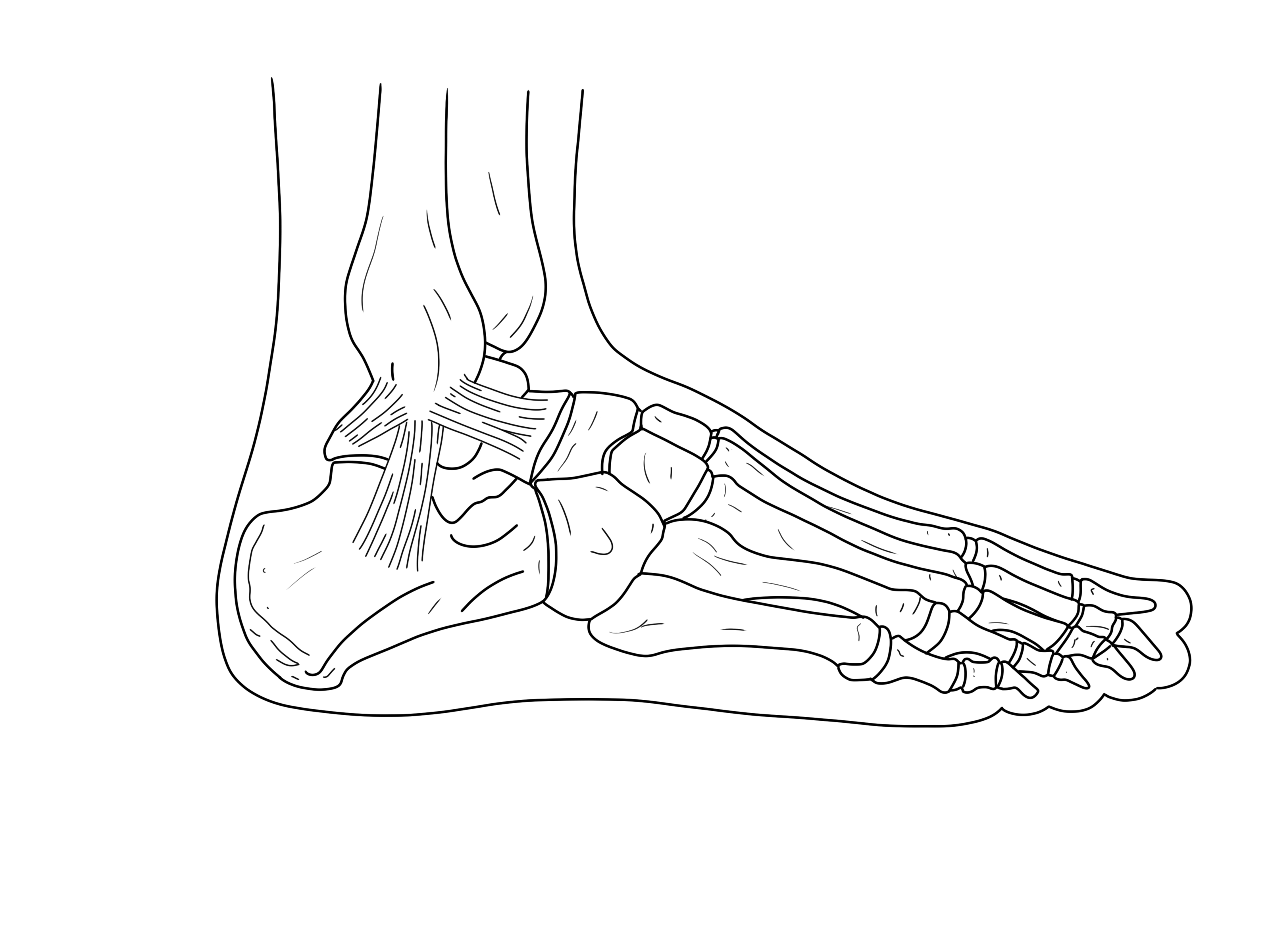
References
Chiha M, Samarasinghe S, Kabaker AS. Thyroid storm: an updated review. J Intensive Care Med 2015.
Douma MJ, Aves T, Allan KS, et al. First aid cooling techniques for heat stroke and exertional hyperthermia: A systematic review and meta-analysis. Resuscitation 2020.
Druyan A, Janovich R, Heled Y. Misdiagnosis of exertional heat stroke and improper medical treatment. Mil Med 2011.
Epstein Y, Yanovich R. Heatstroke. N Engl J Med 2019.
Gaudio FG, Grissom CK. Cooling Methods in Heat Stroke. J Emerg Med 2016.
Gauer R, Meyers BK. Heat-Related Illnesses. Am Fam Physician 2019; 99:482.
Klein Klouwenberg PM, Ong DS, Bonten MJ, Cremer OL. Classification of sepsis, severe sepsis and septic shock: the impact of minor variations in data capture and definition of SIRS criteria. Intensive Care Med 2012.
Pileggi DJ, Cook AM. Neuroleptic Malignant Syndrome. Ann Pharmacother 2016.
Rublee C, Dresser C, Giudice C, et al. Evidence-Based Heatstroke Management in the Emergency Department. West J Emerg Med 2021.
Sorensen C, Hess J. Treatment and Prevention of Heat-Related Illness. N Engl J Med 2022.
Authorship
Infographic Written and Composed by Akanksha Vaishnav, MD, PGY-1 University of Cincinnati Department of Emergency Medicine
Peer Review, Editing and Posting by Jeffery Hill, MD MEd, Associate Professor of Emergency Medicine, University of Cincinnati
Cite As
Vaishnav, A. Hill, J. (April 26, 2023) Acute Hyperthermia in the Emergency Department. TamingtheSRU. https://www.tamingthesru.com/blog/core-content/acute-hyperthermia-in-the-emergency-department

IV Metoprolol vs Diltiazem for A fib with RVR and Concomitant Heart Failure
The management of atrial fibrillation with rapid ventricular response is often complicated by the presence of heart failure with reduced ejection fraction. The presence of HFrEF limits pharmacologic options for rate control. This podcast will cover a retrospective study looking at the use of metoprolol vs diltiazem in patients with A fib with RVR and concomitant heart failure
Background
Atrial fibrillation is a prevalent disease entity that affects over 500,000 new people annually. A subset of these patients develop an accelerated rate either primarily or due to other factors known as rapid ventricular response. If untreated, atrial fibrillation can increase clot formation and lead to an increased risk for heart attack and stroke as well as development of congestive heart failure. Concomitant atrial fibrillation and heart failure makes treatment more difficult and increases the odds of mortality up to 57% compared to isolated heart failure. For treatment, the American Heart Association’s 2019 guidelines recommend limiting use of non-dihydropyridiine CCB due to potential for negative inotropic effects. However, it is still used in certain cases. Currently, there is limited data investigating outcome differences between beta blockers and calcium channel blockers for atrial fibrillation with rapid ventricular response in a patient diagnosed with heart failure.
Study design
This was a retrospective-IRB approved study evaluating adult patients >18 years of age who had documented heart failure with reduced ejection fraction (HFrEF) or heart failure with preserved ejection fractures (HFpEF). Inclusion criteria included echocardiogram performed within previous 12 moths or within 24 hours of admission in patients who presented in atrial fibrillation with rapid ventricular response to a heart rate (HR) >120. Exclusion criteria included those presenting in an exacerbation of their heart failure or those treated with other agents within first 30 minutes of receiving the study drug.
Their primary effectiveness outcome was successful HR control <110bpm within 30 minutes and secondary included successful control at 60 minutes and HR control at ED transfer/discharge. Additional secondary outcomes of interest included evaluation of HR in beats per minute, reduction in HR both numerically and those >20%, time to adequate HR control, total dose of meds give, additional agents used for HR control, use of fluids and crossover to other medications.
Safety outcomes were monitored and included bradycardia, hypotension +/- vasopressor use, dyspnea and hypoxia, change in EF on subsequent echocardiogram and acute kidney injury +/- use of renal replacement therapy.
Results
They screened 2,580 patients and 193 were included (59 for metoprolol, 134 diltiazem). Groups were decently matched overall with average age 78, predominantly white and female. Overall, mostly HFpEF with average EF 48%. They found successful rate control at 30 mins not significantly different with 55% for diltiazem and 41% for metoprolol (p=0.063). Overall, diltiazem was faster, had a grossly higher reduction in HR at 30 minutes and 60 minutes and a greater frequency of reduction >20% at 30 and 60 minutes as well as time of discharge/transfer. There were similar lengths of say overall and no significant difference in safety outcomes.
Subgroup analysis
The larger subgroup was HFpEF (n=123) and overall they found more frequent rate control with diltiazem at 30 mins 56% vs metoprolol 36% (p=0.037). Additionally, a higher HR reduction at 30 and 60 minutes, as well as ED discharge/transfer. However, notably, these patients had a higher frequency of hypotension 28% vs 7% (p=0.005).
For the HFrEF group, there were similar rates of HR control at 30 minutes. The diltiazem group had greater reduction in HR at 30 and 60 minutes. However, no difference noted in success of HR control at 60 min and transfer/discharge. There was no difference in safety outcomes. They did note on echocardiogram evaluation that there was a trend towards improvement in EF with metoprolol group (10% vs 2.1% p=0.05).
Discussion
The researchers concluded that there was no difference in rate control at 30 minutes when utilizing diltiazem vs metoprolol based on this study. Other interesting notes included that those who received metoprolol were more likely to crossover to diltiazem after 30 minutes and additionally received more doses of medication overall. The study is unfortunately limited due to retrospective nature, probable selection bias due to small sample size and has limited generalizability due to exclusion of concurrent heart failure exacerbation on presentation. Additionally, average EF very high overall with predominantly HFpEF patients. Regarding diltiazem, it is worth noting that it has potential to be unsafe, as more hypotension developed overall, but very few patients required pressors. Additionally, many of the patients in the study ended up on diltiazem drips whether it be for ease of use, titratability or other factors. A wonderful next step would be to see a randomized control trial investigating this same clinical question…
Authorship
Written by Bronwyn Finney, MD, PGY-3 University of Cincinnati Department of Emergency Medicine
Peer Review, Editing, Podcast Editing by Jeffery Hill, MD MEd, Associate Professor, University of Cincinnati Department of Emergency Medicine
Cite As
Finney, B. Hill, J. (April 22, 2023) IV Metoprolol vs Diltiazem for A fib with RVR and Concomitant Heart Failure. TamingtheSRU. https://www.tamingthesru.com/blog/2023/4/22/iv-metoprolol-vs-diltiazem-for-a-fib-with-rvr-and-concomitant-heart-failure