Deep space infections
Skin and soft tissue infections are often apparent on physical exam and help narrow your differential and tailor your treatment. However, deep to our dermal layers, subcutaneous tissue, and superficial fascia lie “potential spaces” between fascial planes. When an infection inhabits this area, it is referred to as a Deep Space Infection (DSI). DSI’s can present in a variety of anatomical regions and their management in the Emergency Department focuses on source control and understanding the life-threats they can impose depending on the anatomical planes they spread into.
Background
Objectively speaking, DSI’s do not originate in a true physiological space at all. Normally, fascial layers lie parallel to each other. However, when an infection seeds itself between two facial layers and there is pus formation, this “potential space” expands and the abscess can theoretically extend to the limits that these two fascial planes lie in parallel. Because of this extension and anatomical distinction, DSI’s can be occult and require careful consideration during the workup, treatment, and management of complications in the Emergency Department.
It is important to note that there are more DSI’s than the ones we will focus on in this post (spinal epidural abscess, iliopsoas abscess, osteomyelitis, discitis, etc). For today, we will cover the following:
Deep Neck Space Infections (DNSI)
Deep Space Infections of the Hand and Wrist
Deep Neck Space InfectionS
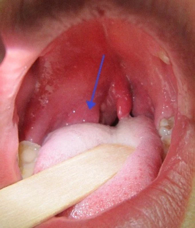
Peritonsillar abscess. Wikipedia. https://en.wikipedia.org/wiki/Peritonsillar_abscess. Accessed September 9, 2026.
Peritonsillar Abscess (PTA)
Epidemiology: The most common DSI of the head and neck; incidence ranges from 30 per 100,000 people per year to 37 per 100,000 people per year and up to 125 per 100,000 people per year ages 14-21 (1)
Etiology: Originally thought to be a sequalae of acute exudative tonsillitis but due to PTA incidence being year-round and tonsillitis mostly seasonal, it is now thought to be due to duct obstruction. Weber glands are a group of salivary glands connected via a duct to the surface of the tonsil. If this duct becomes obstructed and the glands undergo inflammation, cellulitis can progress to abscess formation within the soft palate
Risk Factors:
Peridontal disease and tobacco smoking
Teenagers and young adults aged 14-21
Recent acute tonsillitis or pharyngitis
Presentation:
Constitutional symptoms: Feeling ill, malaised, febrile
Localized symptoms: throat pain (ipsilateral > bilateral and can refer to ipsilateral ear) odynophagia, dysphagia, drooling
Physical Exam:
Trismus: due to inflammation of soft tissue and masticator muscle spasm
“Hot-potato voice” – Have you ever tried to talk with hot food in your mouth? Imagine the mumbling quality of your voice. Patients will have this.
Unilateral cervical lymphadenopathy
Oropharynx: swelling and erythema of anterior tonsil and soft palate superficial to the tonsil
Tonsil displaced inferiorly and medially, contralateral deviation of uvula
Diagnosis:
Mainly diagnosed clinically however imaging modalities can be used
CT Head/Neck with Contrast if:
Concern for infection spreading beyond peritonsillar space
Concern for airway compromise
To confirm presence of abscess
Ultrasound also can be used to confirm or rule out presence of abscess (peritonsillar cellulitis) and avoid unnecessary needle/scalpel intervention
Inflammatory markers can be used to assess severity but will not change ED management
ED Management:
Needle aspiration vs. incision and drainage. Neither are superior so decision lies at discretion of provider comfort (1)
Considerations: Location of the internal carotid artery during drainage. Recent studies suggest actual risk is lower than perceived theoretical risk (2)
Disposition
Discharge (can tolerate PO, pain improved after drainage, airway intact)
Outpatient Antibiotics options (10-14 day course): Amoxicillin/clavulanate OR Clindamycin
Must cover GAS and oral anaerobes
Admit (Inability to tolerate PO, uncontrolled pain, procedure complications, unreliable follow up):
Inpatient Antibiotics options (10-14 day course): Ampicillin/sulbactam OR metronidazole + ceftriaxone OR clindamycin + levofloxacin
If suspect MRSA, add MRSA coverage with vancomycin
Steroids:
Single dose IM or IV steroids can decrease pain and improve oral fluid intake in first 12-24 hours after administration (3)
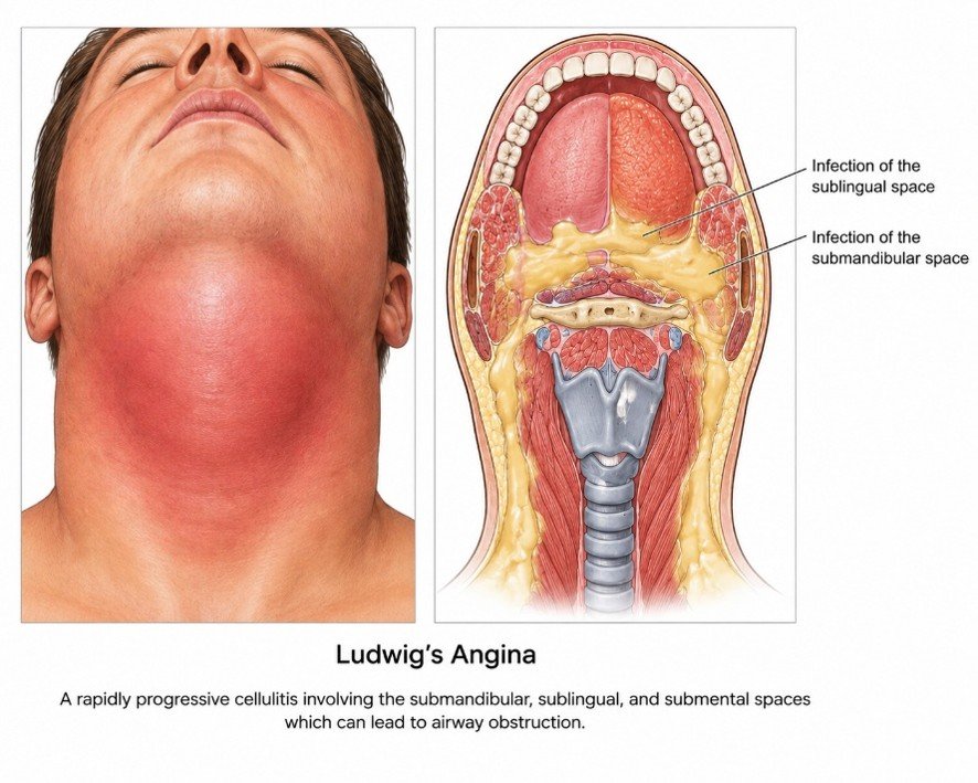
Open AI. (2026, August 24). Ludwig's angina based on the anatomical involvement of the sublingual and submandibular spaces [AI-generated image]. Chat GPT Images.
Ludwig’s Angina
Epidemiology:
Mortality 0.3-8%
47.2% underwent surgical drainage; 3.3% required surgical airway; 4.6% required nonsurgical airway (4)
Etiology:
Rapidly expanding cellulitis of submandibular, sublingual, and/or submental spaces
Odontogenic in 80-89% of cases (2nd and 3rd mandibular molars usually) (5)
Risk factors:
Poor dentition and periodontal disease
Higher risk of airway compromise and necrotizing infections in patient with diabetes as well as those who are immunocompromised
Presentation:
Symptoms: Chief complaint can be “toothache”. Patients report mouth/neck pain, odynophagia, dysphagia, drooling.
Physical Exam:
“Woody”, indurated, tender floor of mouth and can extend to mandible
Edema in floor of mouth can displace tongue posteriorly
Trismus is a late finding
Diagnosis:
Clinical diagnosis but CT Neck/Soft Tissue with contrast can be obtained to determine extent of involvement (but only if patient is safe to leave the ED)
Consider POC U/S for those who cannot tolerate being supine for CT imaging
ED Management:
Definitive management: (1) secure airway and (2) surgical incision and drainage. Evidence is limited but steroids can reduce edema and may reduce risk of airway obstruction
Keep patient upright, supine position worsens obstruction
Multidisciplinary care: Emergent consult to ENT
Securing Airway: Awake fiberoptic nasotracheal intubation is first-line; with surgical airway as backup
Disposition: ICU for close airway-monitoring and IV Antibiotic treatment post-surgical drainage
Retropharyngeal and Parapharyngeal Abscesses (RPA/PPA):
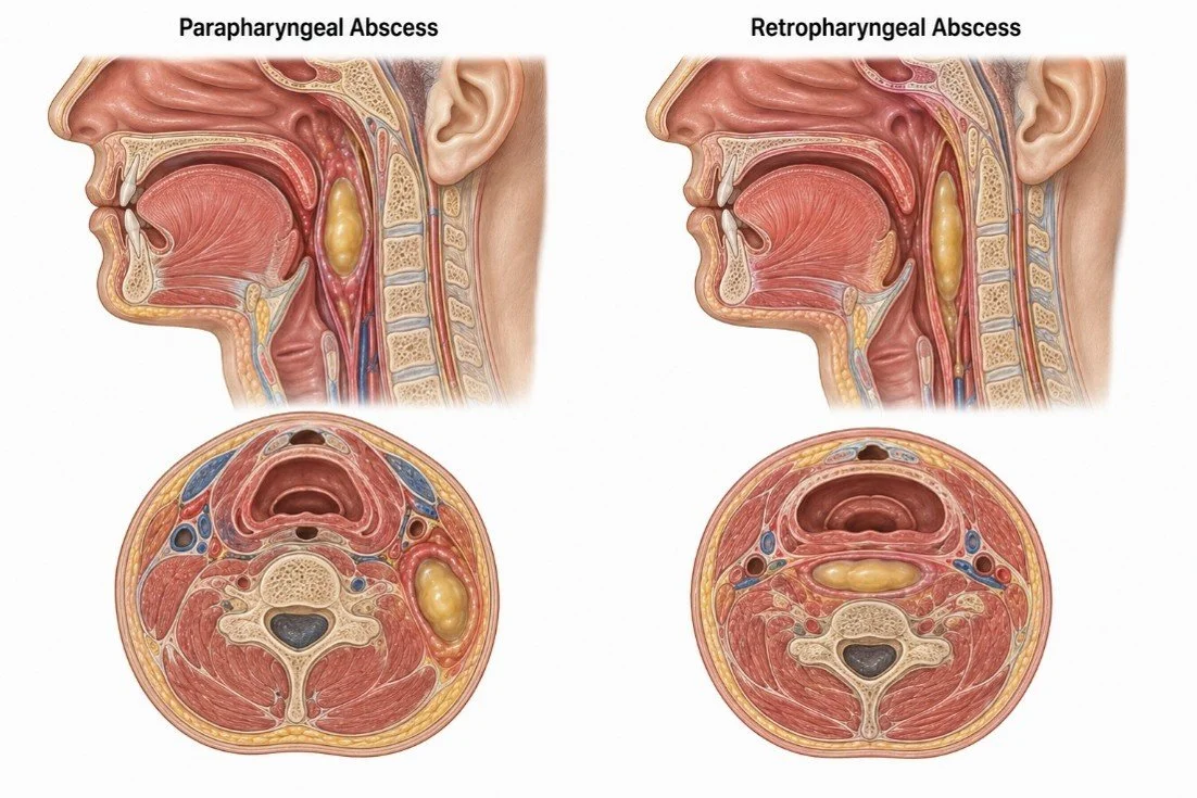
Open AI. (2026, August 24). Retropharyngeal and parapharyngeal Abscess Graphic [AI-generated image]. ChatGPT Images.
Epidemiology: Combined incidence 2.64 per 100,000 people (6)
RPA: pathology of young children mostly (highest in children < 5 y/o) due to suppuration of retropharyngeal lymph nodes peaking at ~ 5y/o
PPA: Second most common DNSI after PTA. Median age is 45 y/o. (6)
Etiology:
RPA:
Children: due to suppuration of prominent retropharyngeal lymph nodes
Adults: pharyngeal trauma or adjacent infection
Risk Factors: young age (2-5; boys > girls), preceding URI
PPA: Most commonly arise from odontogenic infections
Risk factors: odontogenic infection, tonsillar/pharyngeal infection, diabetes, smoking, immunosuppression
But where exactly are they?
RPA: The retropharyngeal space lies directly behind the pharynx and esophagus, anterior is the posterior pharyngeal fascia and posteriorly is the alar llayer of the deep cervical fascia
PPA: Parapharyngeal abscess lies lateral to the pharyngeal constrictor muscle
Presentation:
Symptoms: Fever, odynophagia, dysphagia, drooling, neck pain and stiffness
Of note, manifestation in children can be generalized (fever, poor feeding, irritability) which can lead to misdiagnosis or delayed treatment
Physical Exam:
RPA: neck hyperextension and torticollis
PPA: trismus and medial displacement of the lateral pharyngeal wall or tonsil
Diagnosis:
Imaging: CT Neck with Contrast is first-line
Lateral radiographs and POCUS not reliable for evaluating space involvement
Inflammatory markers
Again, used to assist with triaging and severity rather than diagnosis
Pediatric population: elevated CRP (≥50 mg/L) independently associated with ICU admission (7)
ED Management:
Prioritize airway: This is much less of an issue compared to Ludwig’s angina. While Ludwig’s angina directly displaces the tongue posteriorly providing obstruction at the oropharyngeal and supraglottic level, RPA/PPA provide their obstruction posterior and/or lateral to the pharynx. RPA and PPA also progress more slowly than Ludwig’s typically.
Broad-spectrum IV Antibiotics (coverage and regimen same as PTA)
60% resolve with initial 24-48h of medical management alone (8)
PPA: Penicillin G + metronidazole
Multidisciplinary care: ENT consultation for surgical drainage
Deep Space Infections of Hand and Wrist
Flexor tenosynovitis
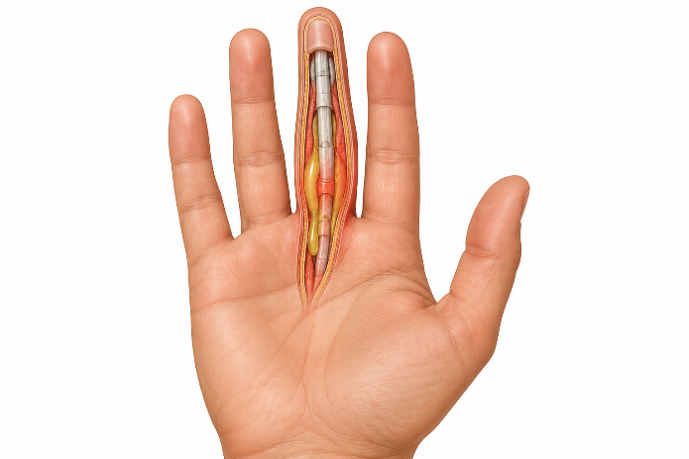
OpenAI. (2026, August 25). Flexor Tenosynovitis [AI-generated image]. ChatGPT Images.
Epidemiology:
Accounts for 2.5-9.4% of all hand infections (9)
Risk Factors: Male sex, older age, comorbidity, and low occupational qualification, diabetes, peripheral vascular disease, renal failure, and immunosuppression
Etiology:
Most common mechanism is penetrating trauma/puncture wound to the volar digit, introducing bacteria along the tendon sheath.
Sheath already poorly vascularized and pus is generated under immense pressure
Can progress within hours with necrosis, adhesions, and digital stiffness. If not recognized, infection can spread into deep spaces of forearm.
Presentation:
Worsening pain, swelling, and stiffness of involved finger following penetrating trauma to volar aspect of finger. Systemic symptoms not always present
Diagnosis
History
Four Kanavel Signs. Not all signs are required
Fusiform “sausage-like” swelling on entire digit
Semi-flexed resting posture of digit
Tenderness along course of flexor tendon sheath
Pain with passive extension of digit (often earliest and most sensitive sign)
Imaging: used to support diagnosis but not confirmatory
POCUS: anechoic/hypoechoic fluid within tendon sheath, sheath thickening, peritendinous edema
Can help distinguish from abscess/cellulitis
Plain radiographs: useful when evaluating for retained foreign body, fracture, subcutaneous gas, or osteomyelitis
MRI: highly sensitive but not appropriate for ED as this should not delay surgical consultation
Inflammatory markers:
Not diagnostic; CRP and ESR have 100% specificity but sensitivity is low (41% and 76%, respectively) (10)
Normal markers do not rule out pathology
ED Management:
Empiric IV Antibiotics
Vancomycin 25-30mg/kg IV loading dose then 15-20mg/kg two to three times daily PLUS (ceftriaxone 2g IV once daily or levofloxacin 750mg IV once daily)
If mechanism is adult bite wound
Ampicillin/sulbactam 3g IV four times daily
Ceftriaxone 2g IV once daily AND metronidazole 500mg IV three times daily
Multidisciplinary care: emergent hand surgery consultation
Immobilize (splint in position of function) and elevate hand
Obtain wound/blood cultures as appropriate
Analgesia, tetanus prophylaxis if warranted
Disposition: Admission for IV antibiotic treatment and operative surgical management
Post by: Blake Martin, MD
Dr. Martin is a PGY-1 in Emergency Medicine at the University of Cincinnati
Editing by: Ryan LaFollette, MD
Dr. Ryan LaFollette is an APD in Emergency Medicine at the University of Cincinnati and Co-editor of Tamingthesru.com
CO-Editing by: Haley Escheman, mph
Haley is a Program Administrator at the University of Cincinnati College of Medicine
References
Chang BA, Thamboo A, Burton MJ, Diamond C, Nunez DA. Needle aspiration versus incision and drainage for the treatment of peritonsillar abscess. Cochrane Database Syst Rev. 2016;(12):CD006287. doi:10.1002/14651858.CD006287.pub4.
Jufara TJ, Duneant E, Nigussie AH, et al. Risk of internal carotid injury during peritonsillar abscess drainage in the emergency department. Am J Emerg Med. 2025;93:132–134. doi:10.1016/j.ajem.2025.04.005.
Chau JK, Seikaly HR, Harris JR, Williams DC, Corsten M, Allen H. Corticosteroids in peritonsillar abscess treatment: a blinded placebo-controlled clinical trial. Laryngoscope. 2014;124(1):97-103.
McDonnough JA, Ladzekpo DA, Yi I, Bond WR Jr, Ortega G, Kalejaiye AO. Epidemiology and resource utilization of Ludwig's angina ED visits in the United States 2006–2014. Laryngoscope. 2019;129(9):2041–2044. doi:10.1002/lary.27734.
Bridwell R, Gottlieb M, Koyfman A, Long B. Diagnosis and management of Ludwig's angina: An evidence-based review. Am J Emerg Med. 2021;41:1–5. doi:10.1016/j.ajem.2020.12.030.
Yang TH, Xirasagar S, Cheng YF, Wu CS, Kao YW, Lin HC. A nationwide population-based study on the incidence of parapharyngeal and retropharyngeal abscess—A 10-year study. Int J Environ Res Public Health. 2021;18(3):1049. doi:10.3390/ijerph18031049.
Kurt F, Yiğit H, Kaynak MO, et al. Predictors of pediatric intensive care unit admission in pediatric deep neck infections: A retrospective observational study. Medicine (Baltimore). 2025;104(48):e46302. doi:10.1097/MD.0000000000046302.
Bochner ER, Gangar M, Belamarich PF. A Clinical Approach to Tonsillitis, Tonsillar Hypertrophy, and Peritonsillar and Retropharyngeal Abscesses. Pediatrics in Review. 2017;38(2):81–92. https://doi.org/10.1542/pir.2016-0072
Forder BH, Hennessy M, Turner B, Wormald J. Systematic review of treatment for pyogenic flexor tenosynovitis of the hand. J Plast Reconstr Aesthet Surg. 2024;96:1-XX.
Bishop GB, Born T, Kakar S, Jawa A. The diagnostic accuracy of inflammatory blood markers for purulent flexor tenosynovitis. J Hand Surg Am. 2013;38(11):2208-2211.Draeger RW, Bynum DK Jr. Flexor tendon sheath infections of the hand. J Am Acad Orthop Surg. 2012;20(6):373–382. doi:10.5435/JAAOS-20-06-373.
Galioto NJ. Peritonsillar Abscess. Am Fam Physician. 2017;95(8):501–506.
Kuppalli K, Livorsi D, Talati NJ, Osborn M. Lemierre's syndrome due to Fusobacterium necrophorum. Lancet Infect Dis. 2012;12(10):808–815.
Lawrence R, Bateman N. Controversies in the management of deep neck space infection in children: An evidence-based review. Clin Otolaryngol. 2017;42:156–163. doi:10.1111/coa.12692.
Mehta P, Thoppil J, Koyfman A, Long B. High risk and low prevalence diseases: Flexor tenosynovitis. Am J Emerg Med. 2024;77:132–138. doi:10.1016/j.ajem.2023.12.023.
Page C, Biet A, Zaatar R, Strunski V. Parapharyngeal abscess: Diagnosis and treatment. Eur Arch Otorhinolaryngol. 2008;265(6):681–686. doi:10.1007/s00405-007-0524-5.
Patterson HC, Kelly JH, Strome M. Ludwig's angina: An update. Laryngoscope. 1982;92(4):370–378.
Sheikh Z, Yu B, Heywood E, Quraishi N, Quraishi S. The assessment and management of deep neck space infections in adults: A systematic review and qualitative evidence synthesis. Clin Otolaryngol. 2023;48(4):540–562. doi:10.1111/coa.14064.
Uchida D, Sekiguchi T, Yamahara K, et al. Diabetic ketoacidosis triggered by retropharyngeal abscess complicated by descending necrotizing mediastinitis. Intern Med. 2026.
Vallée M, Gaborit B, Meyer J, et al. Ludwig's angina: A diagnostic and surgical priority. Int J Infect Dis. 2020;93:160–162. doi:10.1016/j.ijid.2020.01.028.
Wasson J, Hopkins C, Bowdler D. Did Ludwig's angina kill Ludwig? J Laryngol Otol. 2006.
Yealy D, Mohr N, Shapiro N. Early Care of Adults With Suspected Sepsis in the Emergency Department and Out-of-Hospital Environment: A Consensus-Based Task Force Report. Ann Emerg Med. 2021;78:1–19.