
Tamingthesru
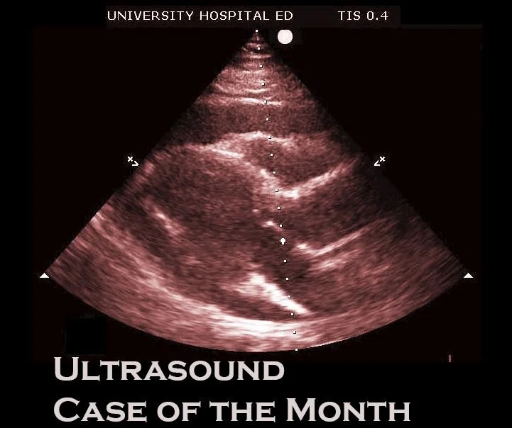
US Case of the Month - Sonography of Shoulders
Shoulder looking off and don’t have / have time to look for an XRay? Join Dr. Kate Connelly as she takes us through the sonographic evaluation of shoulders and real time evaluation of dislocation reductions.
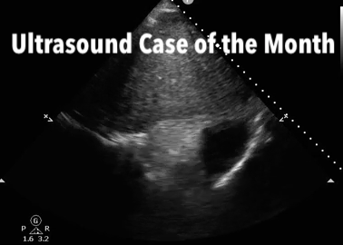
Ultrasound case of the month - Placement comes first
This month, the Taming the SRU ultrasound team details some of the procedural applications of ultrasound in the midst of the COVID-19 pandemic, fresh from two of the minds our intern class: Drs. Hamza Ijaz and Chris Zaleky. This combo post will discuss the use of ultrasound to confirm placement of both endotracheal tubes and central venous catheters.

The Anatomy of Femoral Vascular Access
Prior to the widespread availability of point-of-care ultrasonography, invasive medical procedures were performed by the “landmark method”. Landmark methods are based on surface anatomy, palpation, and sometimes trigonometry, and are fraught with the potential for error. Complications, while unquantified in the misty past, were likely much more common than in the current era of readily available bedside imaging. Vascular access procedures are inarguably safer and more successful when guided by sonography, but interpretation of ultrasound images still requires an understanding of both surface and deeper anatomy to relate the two-dimensional screen image to three-dimensional reality. Further, there are circumstances where either the urgency of the resuscitation, or compromised access to the patient, requires that vascular access be obtained using landmarks rather than real-time imaging. In such cases a detailed understanding of regional anatomy is critical to maximize procedural success and minimize complications.