Welcome to the new Tamingthesru
Same great content - new look - improved search
Interpretation of Hand & Wrist Radiographs
You knew that you'd see at least one patient with a FOOSH (Fall On Outstretched Hand) while working in Minor Care. A nice, thin layer of ice laid down by the "Snow-pocalypse" snow storm that came through last night had already caused several patients to slip and fall.
John, your first patient of the day, a 24 year old rushing to work this morning, slipped coming down the steps outside his house. He tells you he landed on his right hand with his arm extended at the elbow and wrist. He is complaining of pain in the dorsal and radial right wrist. You go to examine him, noticing no obvious deformities, lacerations, or abrasions. On palpation, he has tenderness to palpation in the anatomic snuff box and pain with axial loading of the thumb. Suspecting a scaphoid fracture, you order a wrist x-ray.
While waiting for the film to be developed, you look for a refresher on how to interpret these challenging films.
Interpretation of Ankle Radiographs
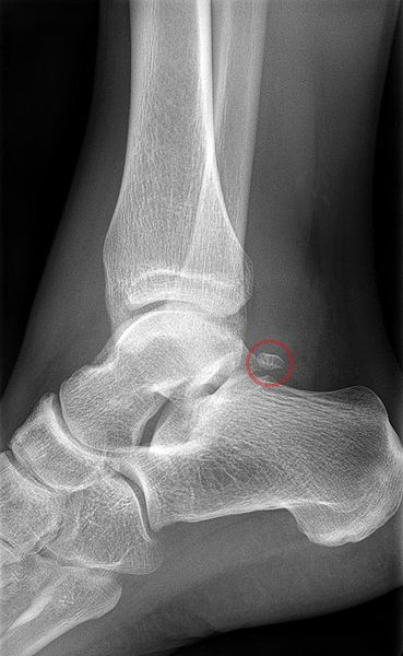
A 43 year old female presents to the ED after "rolling" her ankle while gardening. She states that she was stepping down on a shovel when her ankle rolled. <She describes to you and inversion type injury.> Being a diligent, studious physician, you quickly run through the Ottawa Ankle Rules while you obtain the remainder of you history and physical. She was unable to bear weight immediately after the accident and is, likewise, unable to do so here in the ED. She has no pain with palpation over the medial malleolus but does have significant pain and tenderness with palpation of the lateral malleolus. You quickly decide that this patient will need ankle radiographs to further investigate the possibility of fracture.
But, what views should you order? And, once you get the films back, how do you interpret them. Check out the excellent video embedded below, made by Dr. Claire O'Brien, PGY-1 in the University of Cincinnati Dept. of Emergency Medicine Residency Training program, to find out!
Interpretation of Head CTs
In the video series below, PGY-1 resident, Dr. Gorder, leads us through the key aspects of CT head interpretation. Attention is paid to the development of a rigorous systematic approach to review and interpretation of head CTs to aid in the identification of blood, ischemia, mass, signs of increased ICP, as well as fracture. In the second video, the key anatomic features seen on head CT's are covered.